Showing posts with label subtype. Show all posts

Friday, August 30, 2013

This Month In Blastocystis Research (AUG 2013)

Quite a few papers relevant to Blastocystis research have made it to PubMed over the past month! Therefore, the August version of 'This Month in Blastocystis Research' is more like a list of papers + short descriptions/comments, rather than one or two actual paper reviews.

Dr Aldert Bart and his Dutch colleagues have published a study that confirms data emerging from other parts of Europe. Using microscopy (fixed faecal smears) and PCR, they found an almost 40% prevalence of Blastocystis in returning travelers with symptoms, and a prevalence of 18% in patients referred for other reasons. The distribution of subtypes found in the study population was quite similar to what has been found elsewhere in Europe with ST3 predominating (42%) and the rest of the subtypes attributable to ST1 (22%), ST2 (22%), ST4 (12%), ST6 (1%) and ST7 (1%).

The Tropical Parasitology theme issue on Blastocystis has now gone live. You’ll find a link to the editorial and the three papers included in the symposium here.

In my previous post I referred to a new study from Colombia which includes subtyping of Blastocystis isolates from humans, and a variety of animals, including birds. The paper is interesting for a number of reasons, but first and foremost it confirms the virtual absence of ST4 in humans in S America. Moreover, the study included 70 Blastocystis positive samples from asymptomatic carriers, 40 positive samples from patients with diarrhoea, and 15 positive samples form patients with IBS. Remarkably, all samples from healthy carriers were typed as ST1, those from patients with diarrhoea belonged to ST2, and those from IBS patients to ST3. Such a clear-cut distribution of subtypes across cohorts is unprecedented and of course warrants confirmation and further investigation. In Europe, ST4 is very common in humans, while it appears rare in humans in many other parts of the world. ST4 also appears rare among non-human primates (NHPs), our closest living relatives, and while NPHs and humans otherwise tend to share the same major subtypes (ST1, ST2, and ST3), this suggests that while subtypes 1, 2 and 3 have probably co-evolved with primates, ST4 has only recently entered the primate population with a preference for humans! I have hinted at this many times by now, but I find it extremely interesting which is why I keep repeating it.

There is a paper out by Santos and Rivera from the Philippines comparing microscopy of direct faecal smear with culture and PCR for detection of Blastocystis. They ended up concluding that culture was the best diagnostic modality, but it should be noted that the PCR used in the study targets a 1.8 kbp product (complete SSU rRNA gene!), and much smaller products are usually targeted in diagnostic PCR assays. The Blastocystis real-time PCR developed by me and my colleagues targets a sequence stretch of ~120 bp, securing optimum test sensitivity. The results of the Philippine study should be interpreted with this in mind.

Li et al., have published data on experimental infection of ST1 in Sprague-Dawley rats. Animals belonging to this species appeared susceptible to a ST1 strain isolated from a diarrhoeic patient that had been kept in culture and for which induction of cysts had been performed with a view to infecting the rats. The study confirms that Blastocystis is mainly a parasite of the coecum and colon. The authors found evidence of Blastocystis invasion into the lamina propria in one of the animals, and signs of inflammation in all animals challenged. While it is great to see that experimental models can be sustained and that encystation can be induced in vitro, at least two important factors must be kept in mind to fully comprehend the study: Although cysts were isolated by gradient centrifugation prior to inoculation, it is unlikely that all bacteria have been removed from cyst suspensions; in other words, the cyst preparation is not likely to be 'sterile', and any effect of the potentially accompanying bacterial flora is difficult to determine. Moreover, rats may not be natural hosts of ST1 (very few data available on the topic!), and so, the pathology caused in the rats may be an unlikely finding in humans, who are indeed natural hosts of ST1 and may have developed a high degree of tolerance to this subtype.

Are dogs, wolves, and other canids natural hosts of Blastocystis?

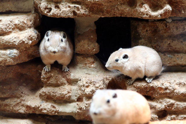
When visiting Australia earlier this month, I had the pleasure of meeting Wenqi Wang and Tawin Inpankaew, both PhD students working at School of Veterinary Science, The University of Queensland Gatton Campus and supervised by Dr Rebecca Traub. One of the foci of this group is to study Blastocystis in animals, for instance in households where animals are kept as pets. Recently, a paper emerged from this group on diversity of Blastocystis subtypes in dogs in different geographical settings, hence domestic/pound dogs from Brisbane, Australia, semi-domesticated dogs from a village in Cambodia, and stray dogs from Mumbai and other Indian cities. Using sensitive PCR methods they observed that almost one fourth of the Indian dogs were infected, while dogs in the Cambodian village and in Queensland remained largely uninfected. Coprophagy and access to Blastocystis-positive stool from different hosts may account for the relatively high prevalence in stray dogs in India, although one might assume that the prevalence would then be even much higher? The team used nested PCR in their study and found four different subtypes in the Indian dogs, including ST1, ST4, ST5 and ST6. Whether all of their data collectively indicate that dogs are not natural hosts of Blastocystis is a matter of debate and remains to be more thoroughly investigated. Indeed, prevalence and subtype data from studies of samples from wild life canids (dingos, jackals, wolves, coyotes, but also foxes and raccoon dogs) would shed further light on this topic.

Finally, for those interested in how Blastocystis deals with oxidative stress and related metabolic issues, there is a paper out on iron-sulphur cluster biogenesis in protozoan parasites by Ali and Nozaki citing works by Tsaousis (2012), Denoeud (2011), Long (2011), and Stechmann (2008).

Literature:

Ali V, & Nozaki T (2013). Iron-sulphur clusters, their biosynthesis, and biological functions in protozoan parasites. Advances in Parasitology, 83, 1-92 PMID: 23876871

Bart A, Wentink-Bonnema EM, Gilis H, Verhaar N, Wassenaar CJ, van Vugt M, Goorhuis A, van Gool T. Diagnosis and subtype analysis of Blastocystis sp. in patients in a hospital setting in the Netherlands. BMC Infectious Diseases, 13:289.

Li J, Deng T, Li X, Cao G, Li X, & Yan Y (2013). A rat model to study Blastocytis subtype 1 infections. Parasitology Research PMID: 23892480 DOI: 10.1007/s00436-013-3536-7

Parija SC (2013). Blastocystis: Status of its pathogenicity. Tropical Parasitology, 3 (1) PMID: 23961433

Parija SC, & Jeremiah S (2013). Blastocystis: Taxonomy, biology and virulence. Tropical Parasitology, 3 (1), 17-25 PMID: 23961437 

Ramírez JD, Sánchez LV, Bautista DC, Corredor AF, Flórez AC, & Stensvold CR (2013). Blastocystis subtypes detected in humans and animals from Colombia. Infection, Genetics and Evolution : Journal of Molecular Epidemiology and Evolutionary Genetics in Infectious Diseases PMID: 23886615

Sekar U, & Shanthi M (2013). Blastocystis: Consensus of treatment and controversies. Tropical Parasitology, 3 (1), 35-9 PMID: 23961439

Stensvold CR (2013). Blastocystis: Genetic diversity and molecular methods for diagnosis and epidemiology. Tropical Parasitology, 3 (1), 26-34 PMID: 23961438  

Wang W, Cuttell L, Bielefeldt-Ohmann H, Inpankaew T, Owen H, & Traub RJ (2013). Diversity of Blastocystis subtypes in dogs in different geographical settings. Parasites & Vectors, 6 PMID: 23883734

Monday, July 29, 2013

Birds of America

Yesterday evening, I was watching another compelling BBC production, broadcast on Danish television: Earthflight, North America. In quite a unique way, the viewers got the rare opportunity to see through the eyes of birds such as eagles, geese, and pelicans and follow birds as they were migrating, escaping, hunting for prey, etc. It made me think of the 19th century masterpiece 'Birds of America' by John James Audubon, which can be viewed in the National History Museum in London. The book features 435 stunning hand-coloured plates that show birds life-size, in natural positions and in their natural habitat.

One of the things that I find interesting - and quite unexplored - is Blastocystis in birds. By 'unexplored' I mean that relatively little sampling has been done, and so the number of observations of Blastocystis in birds is still limited compared to other types of hosts. However, there is a brand new paper out in 'Infection, Genetics and Evolution' which includes observations on Blastocystis in birds (of America!).

You see, I was invited in on a study by colleagues in Colombia who had access to DNA from quite a few faecal samples from a number of host species, including feral birds, and what we found confirms the quite unambiguous trend seen so far: Birds - no matter where on this planet - appear to be colonised mainly by ST6 and ST7. As a matter of fact, in the present study only ST6 was seen in almost 50 Colombian passerine birds of varying species, most of which I believe are limited in geographical distribution to the Americas: Passer domesticus, Thraupis episcopus, Oryzoborus maximiliani, Sicalis flaveola, and Petrochelidon pyrrhonota. Moreover, only one allele of ST6, allele 122, was identified. Notably, the prevalence of Blastocystis in the sampled bird population was 90%. I believe that this is the first official report on Blastocystis in passeriformes. Other major groups of birds previously sampled include galliformes, anseriformes, and ratites (Stensvold et al., 2009; Alfellani et al., 2013).

Other subtypes have been reported in birds (Alfellani et al., 2013), but due to the very low number of samplings these subtypes may be more or less co-incidental/abberant findings. Of note, some samples from birds have been untypable. I have a slight recollection of detecting ST3 in Icelandic rock ptarmigans (in mixed ST infection) collected by Dr Karl Skírnission, but that certainly needs confirmation.

Bird contact/bird droppings - a significant source of Blastocystis in humans? Me feeding some 'Birds of Australia'. Photo by Dr Rebecca J Traub.

ST6 is very rarely seen in humans in Europe. In other parts of the world, for instance in Egypt and some Asian countries, ST6 appears relatively common, but we do not know much about 'bird subtypes' in those particular regions. Also, the situation in the US and Canada is more or less completely unknown (Blastocystis subtyping is something that appears not to attract research groups in North America apart from the one led by Dr Ron Fayer in Beltsville, Maryland).

ST7 is occasionally seen in humans in countries such as Sweden and Denmark. But in my - still limited - experience, individuals infected by these subtypes are not necessarily prone to 'suffer more' from intestinal symptoms than those who do not have these subtypes. While human cases of ST6 (and ST7) may represent cases of zoonotic transmission, it is far to early to draw any conclusions on this. It would be important to compare ST6 and ST7 18S alleles from humans and birds. MLST typing systems for these two subtypes are not yet available, but 18S analysis in itself may prove valuable for molecular epidemiological analyses as in the case of other subtypes (Stensvold et al., 2012).

Walton Ford: "Falling Bough" (Source). You will also see the now extinct Passenger Pigeon in 'Birds of America'.

References:

Ramírez JD, Sánchez LV, Bautista DC, Corredor AF, Flórez AC, & Stensvold CR (2013). Blastocystis subtypes detected in humans and animals from Colombia. Infection, Genetics and Evolution: Journal of Molecular Epidemiology and Evolutionary Genetics in Infectious Diseases PMID: 23886615

Alfellani MA, Taner-Mulla D, Jacob AS, Imeede CA, Yoshikawa H, Stensvold CR, & Clark CG (2013). Genetic diversity of Blastocystis in livestock and zoo animals. Protist, 164 (4), 497-509 PMID: 23770574

Stensvold CR, Alfellani M, & Clark CG (2012). Levels of genetic diversity vary dramatically between Blastocystis subtypes. Infection, Genetics and Evolution: Journal of Molecular Epidemiology and Evolutionary Genetics in Infectious Diseases, 12 (2), 263-73 PMID: 22116021

Stensvold CR, Alfellani MA, Nørskov-Lauritsen S, Prip K, Victory EL, Maddox C, Nielsen HV, & Clark CG (2009). Subtype distribution of Blastocystis isolates from synanthropic and zoo animals and identification of a new subtype. International Journal for Parasitology, 39 (4), 473-9 PMID: 18755193

Friday, June 21, 2013

This Month In Blastocystis Research (JUN 2013)

Another paper in the string of publications coming out from the PhD study by Dr Alfellani (London School of Hygiene and Tropical Medicine) has just appeared in PubMed.

Dr Alfellani and his colleagues have done a great job in analysing a multitude of samples from humans, non-human primates and animals; I have previously blogged about their observations from studies of human and non-human primates. Moreover, they have surveyed available data in order to better discuss their own findings, and the work has contributed significantly to what today is known about the host specificity, genetic diversity, phylogeography and general molecular epidemiology of Blastocystis.

Alfellani's most recent paper is published in the journal Protist, and it deals with the 'Genetic Diversity of Blastocystis in Livestock and Zoo Animals'.

It is quite a large paper which includes a lot of new information and a comprehensive (and hopefully exhaustive) table summarising Blastocystis subtype data in all relevant hosts (humans, non-human primates, other mammals and birds).

I will highlight a couple of things from the paper:

1. Apart from reporting on virtually complete SSU rDNA sequences from a couple of subtypes for which entire SSU rDNA sequences have yet not been available, we also report on three novel subtypes. Until recently, we only knew about 14 subtypes (ST1-ST14), of which ST1-ST9 can be found in humans. Now, three additional subtypes have been identified; ST15 in artiodactyls (camel and sheep) and non-human primates (chimpanzee and gibbon), ST16 in kangaroos, and ST17 in gundis.

The Gundi (Ctenodactylus gundi) is a rodent living mainly in the deserts of Northern Africa. (Source)

2. Novel data arising from analysis of faecal samples from humans and animals in Sebha, Libya, strongly indicate that humans and animals in this area are infected by different subtypes: Humans appear to carry ST1, ST2, and ST3, while synanthropic animals (artiodactyls in this case) mostly have ST5 and ST10 infections, suggesting that livestock is not a major contributor to human Blastocystis infection.

To this end, there is growing evidence of quite a substantial degree of host specificity of Blastocystis.  Even when subtypes overlap between humans and animals, we have accumulating evidence that the strains found in humans and animals are different. This means that the hypothesis that animals constitute an important reservoir of human Blastocystis infections currently has very limited support. It is my clear impression that when a strain of ST6 or ST8 is detected in humans, this strain has most probably been transmitted from an animal source. But we very rarely see these subtypes in humans, at least in Europeans.

It will be extremely interesting to see how the universe of Blastocystis subtypes unfolds... by genetically characterising strains in humans and non-human hosts, we are building up a clearer picture of transmission patterns and evolutionary biology, including our adaptation to Blastocystis, and the parasite's adaptation to us and other hosts.

It is noteworthy that we are starting to see different subtypes in rodents. We have previously thought that generally, rodents were infected by ST4. But now we know that many rodents are not infected, and we also know that rodents may harbour subtypes other than ST4.

So,17 subtypes of Blastocystis are now known. We have probably only seen the top of the iceberg, since many host species have not yet been sampled from, and it is likely that we will see quite a few STs being identified in the nearest future. To this end it is necessary to have a consensus regarding the identification of novel subtypes. Along with the Protist paper we have uploaded a supplementary file (Appendix A, TXT format) with aligned reference sequences that can be used for phylogenetic analysis,  hoping that it will be useful to our colleagues. In a future blog post I will try to address the issues of identifying new subtypes more specifically.

ST15 is one of the more interesting subtypes since it appears to have quite a low host specificity - infecting both non-human primates and artiodactyls. Yet, we have come across it only now. ST15 and ST17 are remarkable in the way that they appear to be closer related to herptile and arthropod lineages, respectively, than to lineages from mammals.

Please note that virtually complete sequences of ST10, ST13, ST14, ST15, and ST17 analysed in the study have been released in GenBank just now.

Further reading:

Alfellani MA, Taner-Mulla D, Jacob AS, Imeede CA, Yoshikawa H, Stensvold CR, & Clark CG (2013). Genetic Diversity of Blastocystis in Livestock and Zoo Animals. Protist, 164 (4), 497-509 PMID: 23770574

Alfellani MA, Stensvold CR, Vidal-Lapiedra A, Onuoha ES, Fagbenro-Beyioku AF, & Clark CG (2013). Variable geographic distribution of Blastocystis subtypes and its potential implications. Acta Tropica, 126 (1), 11-8 PMID: 23290980

Alfellani MA, Jacob AS, Perea NO, Krecek RC, Taner-Mulla D, Verweij JJ, Levecke B, Tannich E, Clark CG, & Stensvold CR (2013). Diversity and distribution of Blastocystis sp. subtypes in non-human primates. Parasitology, 140 (8), 966-71 PMID: 23561720

Thursday, May 9, 2013

YouTube Video on Blastocystis Subtyping

For those who want to venture into Blastocystis subtyping - the easy way - I've recorded and uploaded a video on YouTube fyi.


For even more information, please visit a selection of relevant blog posts here.

Monday, April 29, 2013

'Invasive Blastocystis' in ECCMID 2013

ECCMID - the annual European Congress of Clinical Microbiology and Infectious Diseases (hosted by ESCMID) is currently taking place in Berlin. This year, I'm not attending, but I've been scanning the abstract book for 'Blastocystis', and it appears that an oral presentation was scheduled for yesterday in the "Emerging Infectious Diseases" section:

First of all: it's great to see fellow researchers screening larger (i.e. hundreds) of faecal DNAs by PCR for Blastocystis. I wish more people would do that to produce reliable data on prevalence and subtypes.

Now, as I've already mentioned, there are currently mainly two methods in use for subtyping, barcoding and STS PCR, and recently I evaluated these. To cut a long story short, barcoding is recommended for subtyping, since the STS method, which was used in the study by Tarasova et al. (abstract), appears to miss the majority of ST4 strains (the major genotype), and moreover, no STS primers exist for ST8 and ST9 (or any of the other 8 subtypes identified to date, but which have only been found in animals). So, the subtype data found in this study should be interpreted with this in mind.

Importantly however, I'm not sure whether the authors used the original Yoshikawa STS terminology or the terminology acknowledged in our 2007 consensus.

First, let us assume that consensus terminology is used. Then it's surprising to find ST5 in human samples in the first place, and finding a ST5 prevalence of 45% in a cohort of humans included in a larger study like this is very unlikely based on current evidence of more than 3,000 observations from all over the world, where the overall prevalence of ST5 in humans is <1%. Also, finding so much ST6 is also really striking. Also, if the consensus terminology is used, then I'm a bit puzzled why the authors put emphasis on ST7 not being found, since ST7 is relatively rare in humans.

And so let us assume that consensus terminology was not used, and the original Yoshikawa terminology was used instead. This would translate into STs 4, 6, and 7 not being detected in the CVH group. Which makes sense, since ST6 is extremely rare (at least in Europe), ST7 is only seen on occasion, and, as I said, the majority of ST4 infections are likely to go undetected by the STS method. However, ST4 appears quite common in Europe, and I suspect that it should be quite common in St Petersburg as well. But then there is one thing that comes to my mind: If ST4 infections are common, then there should be a relatively large number of samples detected by PCR which were untypable by PCR...and there is no information on untypable positive samples in the abstract...
But what is more:  STS subtype 5 translates into ST2 in consensus terminology, and similarly STS subtype 6 equals ST5 (yes, it may seem confusing, but we have provided a table in the 2007 consensus paper to make this easy). This means that no matter which of the two terminologies were used, ST5 is seen in abundance in patients with CVH in St Petersburg! Which is a very remarkable observation, and maybe more interesting than the rest of the data, which  I, by the way, find a bit difficult to follow (I expected to learn something about Blastocystis invasion, when I read the title of the abstract, but there is no data or information on invasiveness... and I'm very curious as to how the authors managed to obtain such a high number of samples from 'healthy people'! To evaluate the prevalence of Blastocystis in the control group, demographic data are needed, and a prevalence as low as 5.3% among healthy individuals makes me suspect that this control group consisted of newborns/toddlers who generally have a low prevalence of Blastocystis). Also, since when was ST1 'zoonotic'?

Anyway, often conference abstract are previews of upcoming articles, and so I expect that there will be a paper out soon from this group, and hopefully these issues will be clarified. The occasional confusion in Blastocystis epidemiology could be reduced to a minimum if everyone got into using barcoding and the Blastocystis 18S subtyping site (and go here for a video introduction to Blastocystis subtyping).

Are some citizens of St Petersburg infected by Blastocystis sp. ST5, a subtype seen primarily in livestock and African apes? Source

References:
Tarasova E, Suvorova M, Sigidaev A, Suvorov A. Blastocystis invasion in patients with chronic viral hepatitis in Saint Petersburg. ECCMID 2013 abstract O338.

Alfellani MA, Stensvold CR, Vidal-Lapiedra A, Onuoha ES, Fagbenro-Beyioku AF, & Clark CG (2013). Variable geographic distribution of Blastocystis subtypes and its potential implications. Acta Tropica, 126 (1), 11-8 PMID: 23290980

Stensvold CR (2013). Comparison of sequencing (barcode region) and sequence-tagged-site PCR for Blastocystis subtyping. Journal of Clinical Microbiology, 51 (1), 190-4 PMID: 23115257

Stensvold CR, Suresh GK, Tan KS, Thompson RC, Traub RJ, Viscogliosi E, Yoshikawa H, & Clark CG (2007). Terminology for Blastocystis subtypes--a consensus. Trends in Parasitology, 23 (3), 93-6 PMID: 17241816

Tuesday, April 9, 2013

Blastocystis in Non-Human Primates

If my recent blog post "Blastocystis aux Enfers" could be described as "Blastocystis meets Dante Alighieri", then this post might come across as "Blastocystis meets Sir David Attenborough" (with all due respect to both of these gentlemen!).

Non-human primates (NHPs) include apes (hominoids), Old World monkeys (cercopithecoids), New World Monkeys (ceboids) and prosimians such as lemurs. I have been so fortunate to be involved in a study of Blastocystis in NHPs; a study which was led by Dr Alfellani with several co-investigators, and which has just appeared online in the journal Parasitology (click here to be diverted to the the website - first view article section).

The study is the first of its kind aiming to provide a substantial insight into the host specificity of Blastocystis in NHPs and included subtype observations for 441 captive and free-living animals representing no less than 30 genera; most of the data were generated during the study, while sporadic observations from similar studies were also included.

It was a huge study with a lot of interesting information, and I will try and summarise some of the points here.

Apes such as bonobos, chimps, gorillas and orangutans were colonised by some of the most common subtypes in humans, namely ST1, ST2, and ST3, accounting for about 77% of the cases. Contrary to humans though, ST5 also appeared rather common, accounting for about almost 14% of the cases, and some of the gibbons studied had ST8. Interestingly, a chimp and a gibbon were found to be hosts of a new subtype, ST15.

Old World monkeys were studied to an even larger extent, and again, ST1, ST3 and ST2 predominated, accounting for about 95% of all cases of single subtype infection. Here ST5 was also seen (2%) but only in langurs/lutungs and vervet monkeys. Interestingly, ST8 was seen only in 1/226 cases. ST13 was found by colleagues in Tanzanian colobus monkeys (Petrasova et al., 2011), and 8% of the 226 cases represented mixed/unknown subtype infections.

Woolly monkey (Lagothrix lagotricha) (Source)

New World monkeys included in the study were mainly represented by woolly monkeys, and these were colonised first and foremost by ST8 (49%), but ST3, ST2, ST1 were also found. So was a single case of ST4, which in general appears to be surprisingly rare among NHPs.

A few observations on lemurs were included, and such animals appear to host a vast variety of subtypes with no particular predilection, hence ST1, ST2, ST4, ST8, ST10 and ST15.

Ring-tailed lemur (Lemur catta) (Source).

The most striking differences between humans and NHPs in terms of colonisation by Blastocystis subtypes is that humans are very rarely colonised by ST5, while this subtype appears common in apes and Old World monkeys. ST8 was seen only in arboreal apes and in woolly and howler monkeys, which are also tree-dwellers, and it is tempting to think that ST8 is found mainly in tree-dwelling NHPs; to my knowledge, ST8 has not been found in non-primate hosts, except for once in a bird. Human colonisation by ST8 has been demonstrated only very rarely, for instance in a Danish woman returning from holiday in Indonesia and in animal keepers. Conversely, ST4 is seen extremely rarely in NHPs, while very common in humans in some parts of the world, apparently especially in Europe. These clear discrepancies in subtype distribution in humans and NHPs may boil down to host specificity and/or apparent geographically restricted range of some subtypes.

Another striking observation was that cryptic host specificity exists in ST1 and ST3, meaning that ST1 and ST3 strains found in NHPs overall differ genetically from strains found in humans belonging to the same subtypes, adding support to our previous findings.This suggests that humans are generally colonised by other strains than those found in NHPs. It will be interesting to see, whether other types of hosts sharing these subtypes carry distinct, host-specific strains. While MLST is probably the best way of testing for this, a lot of information can be obtained simply by barcoding. Pets, for instance, may share subtypes seen in humans, and so barcoding of "pet blasto" may be one of the very interesting pathways to knowledge.

We found no evidence of those subtypes that we have nicknamed "avian subtypes", namely ST6 and ST7. In some parts of the world, these two subtypes do not appear uncommon in humans; in Denmark and Sweden, for instance, ST7 is seen on quite a few occasions. But, interestingly, both STs are apparently absent in NHPs.

Langurs - the front cover of one of my favourite books showcasing works by the magnificent Walton Ford.

Incidentally, there is a sequence in GenBank from a gorilla (JX159284) which possibly represents a novel subtype, which is related to reptilian Blastocystis, and so it appears that the host spectrum and diversity of Blastocystis in NHPs continues to unfold.

A recent study saw that faecal microbiomes of wild non-human primates co-vary with host species, hence reflecting host phylogeny. This was evidenced by higher intra-species similarity among wild primate species, which may reflect species specificity of the microbiome in addition to dietary influences. This may in part explain the differences in Blastocystis subtypes seen in different NHP host species, but it is also possible that differences in subtypes reflect differences in habitat (and thereby possibly exposure) or geographical differences in subtype distribution. Indeed Homo sapiens is host to a variety of subtypes, and while ST4 is common in Europe, it appears virtually absent in many other parts of the world. Likewise, the differences in the prevalence of ST8 may reflect differences in geographical distribution, habitat and diet (arboreal vs. ground) as well differences in host specificity.

The overall interesting thing here is the schism of exposure vs. host specificity.


Suggested reading:

Alfellani, M., Jacob, A., Perea, N., Krecek, R., Taner-Mulla, D., Verweij, J., Levecke, B., Tannich, E., Clark, C., & Stensvold, C. (2013). Diversity and distribution of Blastocystis sp. subtypes in non-human primates Parasitology, 1-6 DOI: 10.1017/S0031182013000255

Yildirim S, Yeoman CJ, Sipos M, Torralba M, Wilson BA, Goldberg TL, Stumpf RM, Leigh SR, White BA, & Nelson KE (2010). Characterization of the fecal microbiome from non-human wild primates reveals species specific microbial communities. PloS one, 5 (11) PMID: 21103066

Stensvold CR, Arendrup MC, Nielsen HV, Bada A, & Thorsen S (2008). Symptomatic infection with Blastocystis sp. subtype 8 successfully treated with trimethoprim-sulfamethoxazole. Annals of tropical medicine and parasitology, 102 (3), 271-4 PMID: 18348782

Stensvold CR, Alfellani M, & Clark CG (2012). Levels of genetic diversity vary dramatically between Blastocystis subtypes. Infection, genetics and evolution : journal of molecular epidemiology and evolutionary genetics in infectious diseases, 12 (2), 263-73 PMID: 22116021

Saturday, February 2, 2013

Blog Feedback

I'm very thankful for all the positive feedback I get from readers across the globe, mostly by email. Due to time limits I can only respond to 5-10% of the mail, and I'm sorry for not getting back to the rest of you.

Meanwhile, this blog currently holds more than 60 posts, and you will also find a lot of key words in the right side bar, so take your time and browse a few posts or look up a few relevant key words, -  you might find an answer to one or more  of your questions.

Having said that, I try to read all my email, and I am listening! The feedback and questions that I get are vital for our work and help us identify the avenues that we need to take to unveil the many mysteries of Blastocystis.

And let me just say this for now: A proper microbiological work-up (by state-of-the-art methods, including PCR for intestinal parasites), is something that is offered on a routine basis in only very few laboratories, and also the number of clinically orientated Blastocystis research centres can be counted on one hand, I believe. Subtyping of Blastocystis is currently done mostly in epidemiological surveys (as part of research projects), and I suspect that our lab is one of the very few labs in the world doing subtyping on a routine basis.

Oh, and I've been asked by some readers about how to get blog updates. It's easy: You can follow this blog by email, - just scroll down and find "follow by email" in the right side bar and enter your email address. You can also subscribe to posts via atom (go to the very bottom of the page).

And then here's a little something about stomach acidity and intestinal microbiota from Scientific American, - but make sure to read the comments underneath the post too!
 

Monday, January 14, 2013

A Penny For Your Thoughts

So, what should we do about Blastocystis? What do we want to know?

I believe the imminent answer to the latter question is easy: We want to know whether it’s pathogenic, whether we should treat it and how. But I also think that there are many other interesting aspects of Blastocystis which are also of broad interest to the general public, namely: How about the many cases of asymptomatic Blastocystis carriage? What does Blastocystis do in our guts? Could it have any potentially beneficial impact on our health?

Given the fact that Blastocystis has not been implicated in any outbreaks (admittedly: I guess that no one actually ever looked for Blastocystis in outbreak investigations... except for me!), I reckon that the chance of it being involved in acute diarrhoea is small. So, in that respect it's very different from the other intestinal protists such as Giardia, Cryptosporidium, Cyclospora, microsporidia, even Entamoeba histolytica. It's actually more reminiscent of helminth infections, which are are often chronic, and when light hardly give rise to symptoms (depending on species that is!).So I'm more thinking along the lines of co-evolution, adaptation, etc.

Maybe future research will call for a shift in paradigm, but until then I think that we should do what we already can, just at a larger scale and see where it takes us, namely:

Saturday, January 5, 2013

Where Are We On Blastocystis Subtypes?

As mentioned, Blastocystis exhibits remarkable intrageneric diversity, which is continuously being explored by us and our colleagues. We are convinced that the genus of Blastocystis comprises multiple species, but for now we call them "ribosomal lineages" or "subtypes" and allocate numbers to each subtype, hence ST1, ST2, etc. While the number of subtypes that can be found in humans remains stable, we and our colleagues are still expanding the subtype universe in non-human hosts (I will be blogging on this shortly).

Barcoding currently represents state-of-the-art in Blastocystis subtyping, and luckily this method appears to gain a foothold in labs across the world.

Nine subtypes have been found in humans, but some of them only on rare occasions. A recent study going out from London School of Hygiene and Tropical Medicine and led by Dr Alfellani and published just now in Acta Tropica looked at 356 Blastocystis sequences from samples from the UK and Libya, but also from sub-Saharan Africa, namely Liberia and Nigeria.


Saturday, August 18, 2012

To Treat or Not To Treat... But How?

In the "To Treat or Not To Treat" series (please look up previous post here), we have come to the "...But How?" episode.

Blastocystis may be susceptible to a number of drugs - in vitro. In vitro is not the opposite of in vivo. In vitro just  means that the test has been done on an organism that has been isolated from its usual habitat and tested e.g. in a flask, test tube, etc. In the lab, strains can be challenged and manipulated in multiple ways, but there is no guarantee that the outcome of an in vitro susceptibility test is reproducible in vivo, i.e. when the organism is challenged in its natural habitat and under "natural" conditions. Hence, if you test Blastocystis against metronidazole or any other compound (such as iodine) in vitro, and you observe an effect, you cannot rely on being able to reproduce the effect in vivo. This is due to a variety of reasons including pharmaco-kinetics and pharmaco-dynamics, including the ability of the drug to reach the parasite in its ecological niche, impact of the drug on other micro-organisms, drug interactions, strain-dependent differences in susceptibility (including inherent or acquired resistance), etc.

We recently described a case in which a woman with irritable bowel syndrome (according to the Rome III criteria) had both Blastocystis subtype 9 (ST9) and Dientamoeba fragilis. In order to try and eradicate the parasites and to see whether any eradication would impact on her clinical situation, she received multiple courses of antibiotic treatment:

1. Metronidazole (750 mg x 3/d for 10 days)
2. Tetracycline (500 mg x 4/d for 10 days)
3. Trimethoprim + Sulfamethoxazole (TMP 800 mg + SXT 160 mg x 2/d for 7 days)
4. Mebendazole + Metronidazole (100 mg x 2 separated by 2 weeks; subsequently metronidazole as in 1.)
5. Paromomycin + Metronidazole (PM 500 mg + MZ 170 mg x 3/d for 10 days)

Mebendazole was given to the entire household due to suspicion of pinworm infection running in the family that could be a potential reservoir of D. fragilis (re-)infection.

No clinical alleviation was seen throughout this period.

PCR-based detection of Blastocystis and D. fragilis was used to evaluate  faecal samples 5-10 days post-treatment: Microbiological effect was seen only on D. fragilis which was cleared only after treatment with PM + MZ (5).

So, Blastocystis "survived" this series of antimicrobial treatment. In Denmark, no further relevant treatment options are available for general use (actually, even the use of Humatin (PM) needs a special license).

None of the patient's family members or pets were found to be colonised by the same strain, probably indicating that there was no "local" reservoir for ST9, and that the repeated finding of ST9 was not due to re-infection.

It may be so that Blastocystis requires a certain intestinal bacterial flora to establish. However, we expect that substantial perturbations in the intestinal flora must have taken place during the patient's various treatments, and therefore Blastocystis must be able to quickly overcome and adapt to such perturbations. It may add to the conundrum that in this case the woman harboured ST9, which is only very rarely seen in humans, and we might therefore deduce that its presence would be more volatile. No animal/environmental reservoir has yet been identified for ST9.

There is no doubt that microbiomic profiling of the intestinal flora would be of great benefit in a case like this. If data could be achieved on the impact of these drugs on the relative bacterial structure and function by metagenomic approaches, then this would allow us to explore the changes in the general flora that Blastocystis is capable of withstanding. Certainly, none of these drugs had a measurable in-vivo protistocidal effect on Blastocystis when administered as shown.

I re-emphasise that it is far from certain that Blastocystis is capable of inducing disease, directly or indirectly, and hence, we do not know if, and in which situations, we should aim at eradicating it. Suffice it to say, that in our hands and with the compounds that are available for general use in Denmark, it is apparently extremely challenging to eradicate Blastocystis, if at all possible.

Microbe Resilience (Source)

Further reading:

Coyle CM, Varughese J, Weiss LM, & Tanowitz HB (2012). Blastocystis: to treat or not to treat... Clinical infectious diseases : an official publication of the Infectious Diseases Society of America, 54 (1), 105-10 PMID: 22075794

Engsbro AL, & Stensvold CR (2012). Blastocystis: To Treat Or Not To Treat...But How? Clinical infectious diseases : an official publication of the Infectious Diseases Society of America PMID: 22893582

Stensvold CR, Smith HV, Nagel R, Olsen KE, & Traub RJ (2010). Eradication of Blastocystis carriage with antimicrobials: reality or delusion? Journal of clinical gastroenterology, 44 (2), 85-90 PMID: 19834337

Friday, August 10, 2012

Is This A New Subtype?

To quote one of my colleagues attending the recent IWOP 2012 meeting in Tarrytown, NY, Blastocystis subtyping in humans and animals is becoming 'trendy', and so we keep trying to advocate for a standardisation of the metholodology of Blastocystis subtyping.

We recently changed the title of our page at www.pubmlst.org/blastocystis so that now it is called Blastocystis Subtype (18S) and Sequence Typing (MLST) Databases, and we added some text to front page:

In terms of genetic markers, the barcode region (Scicluna et al., 2006) is by far the best represented in publicly available sequence databases, and the correct subtype can be identified by BLAST analysis in the sequence database at the present site. Blasting against this database has the added advantages, compared to using GenBank, of automatically assigning allele types to the SSU-rDNA as well as using the consensus subtype nomenclature (unlike GenBank where the subtype is included only if one was part of the accession submission and no attempt to impose a standard nomenclature is made). In case the sequence does not match any of the ones in the database despite full coverage of the region, this indicates that the sequence represents a new allele or maybe even a new subtype depending on the amount of variation. If a new subtype is suspected, we suggest doing PCR and sequencing of the complete SSU rRNA gene with subsequent phylogenetic analysis using reference sequences.

Now, the last bit is extremely important. We have seen examples of researchers (including ourselves!) assigning sequences to a new a subtype in the absence of complete SSU rDNA data (in fact complete sequences for ST10-ST14 are not yet publicly available!). Doing so has a least two major limitations/drawbacks: Far from all SSU rDNA regions have been validated as being representative of the whole SSU rRNA gene in terms of phylogenetic analysis, and therefore phylogenetic inferences based on non-validated regions may have little or at least less support than anticipated. Moreover, if someone analyses e.g. position 600-1600, and phylogenetic analysis based on this region reveals a potentially new subtype, this makes it impossible for his/her colleague who has data covering positions 1-600 from a Blastocystis isolate that may also represent a new subtype to ascertain whether it might be same subtype (see example below)!

Obtaining complete SSU rDNA sequences directly from faecal DNA may be a cumbersome task but is sometimes possible by combining sequence-specific primers with low-specificity primers such as the RD5 and the RD3 primers (Clark, 1997). If a cultured isolate is available, obviously this makes complete SSU rDNA sequencing much easier.

While it appears that the number of subtypes occurring in humans stays around 9, our gut feeling is that we are yet to uncover quite a few subtypes colonising non-human mammals, and it's great to see an increasing number of teams exploring the genetic diversity of Blastocystis. For instance, Dr Ronald Fayer and his group recently published exciting data on a new Blastocystis subtype in cattle, which they named ST14 (Fayer et al., 2012).

Importantly, caution should be taken to avoid creating confusion in subtype terminology. Confusion can arise when independent researchers assign the same new subtype name (e.g. ST14, ST15, etc.) to novel sequences which in fact belong to different ribosomal lineages, or when incomplete SSU rDNA sequence data are used; this situation was seen recently, when Petrasova et al. (2011), assigned a Colobus sequence to ST5, although it was in fact a ST13 sequence (Clark et al., in press); the situation arose, since Petrasova et al. (2011) did not have data covering the region currently available for ST13 (Parkar et al., 2010), and therefore believed that their sequence was a unique ST5 variant. As for ST14, less than 500 bp are currently available, and these 500 bp are not in the barcode region, making it difficult for all teams using barcoding to compare their data. And so we would like to advocate for making complete SSU rDNA sequences publicly available (Genbank) for potentially new subtypes, for at least two reasons:

1. Phylogenetic inferences based on the complete SSU rDNA will be more robust than those obtained from analysing shorter sequence streches.

2. Complete seqeunces are needed for reference since subtype screening typically includes a single round PCR such as barcoding (Scicluna et al., 2006) amplifying about 550 bp; in the situation where complete SSU rDNAs are available for all known subtypes, it will be quick to analyse, whether a sequence may represent a new subtype, since this will be independent on the SSU rDNA region studied.We therefore hope that complete SSU rDNA sequences will soon be made publicly available for ST10-ST14.

So, when does a complete SSU rDNA sequence represent a new subtype? Well, we have a review paper in press in Advances in Parasitology on recent developments in Blastocystis research, which will be published in less than six months probably, and which also touches on this topic; once the paper is published, I will try and make a summary our thoughts on this...

Further reading:


Clark CG (1997). Extensive genetic diversity in Blastocystis hominis. Molecular and biochemical parasitology, 87 (1), 79-83 PMID: 9233675

Fayer R, Santin M, & Macarisin D (2012). Detection of concurrent infection of dairy cattle with Blastocystis, Cryptosporidium, Giardia, and Enterocytozoon by molecular and microscopic methods. Parasitology research PMID: 22710524

Parkar U, Traub RJ, Vitali S, Elliot A, Levecke B, Robertson I, Geurden T, Steele J, Drake B, & Thompson RC (2010). Molecular characterization of Blastocystis isolates from zoo animals and their animal-keepers. Veterinary parasitology, 169 (1-2), 8-17 PMID: 20089360

Petrášová J, Uzlíková M, Kostka M, Petrželková KJ, Huffman MA, & Modrý D (2011). Diversity and host specificity of Blastocystis in syntopic primates on Rubondo Island, Tanzania. International journal for parasitology, 41 (11), 1113-20 PMID: 21854778
 
Scicluna SM, Tawari B, & Clark CG (2006). DNA barcoding of blastocystis. Protist, 157 (1), 77-85 PMID: 16431158

Friday, June 29, 2012

On Blastocystis and Animal Models

I was recently encouraged by one of my readers to do a blog post on Blastocystis and animal experimental models. This is not exactly my core competence, which probably boils down to the fact that animal models have only been scarcely used in Blastocystis research for reasons that I will try to account for below.

Animal models (mice, rats, guinea pigs) have often been used to study interactions between hosts and microbes as well as the effect of chemotherapeutic interventions. Therefore, one might assume that animal models are an obvious way of potentially establishing a link between Blastocystis and pathology. But currently, the rationale for carrying out some types of Blastocystis experiments on, say, mice or rats is limited. Why? Well, first and foremost because of at least three major issues.

1) Lack of correlation between in vitro and in vivo evidence. Experimental infections of laboratory mice (Elwakil and Hewedi, 2010) resulted in tissue invasion - something never reported in humans. Another study showed increased oxidative stress in Blastocystis infected rats (Chandramathi et al., 2010), again something not linked to human colonisation. Studies that provided evidence for induction of cytokines, contact mediated apoptosis, and barrier disruption all used axenic Blastocystis and in vitro mammalian cell cultures with no evidence that these effect occur in vivo.

2) Host specificity. Blastocystis exhibits extreme genetic diversity and multiple, genetically very different variants (species, subtypes) exist. These subtypes exhibit moderate host specificity. This means that some subtypes are common in one type of host, whereas other subtypes are common in other types of hosts. For instance, ST5 is very common in pigs, but we rarely see it in humans. ST4 is common in rodents, and in some human populations (mainly Europe it seems), but otherwise extremely uncommon. And so on. This means that some subtypes may be difficult to establish in experimental animals. It also means that any pathology detected in the animal, may not be “reproducible” in another host, - maybe due to the fact that this host has adapted to this particular subtype or even strain. Blastocystis is common in a huge variety of animals, and different animals may have adapted do different subtypes. It is not unlikely that this is due to co-evolution, and therefore it may not turn out to be a big surprise if Blastocystis per se is not usually directly associated with disease. It may still be so, however, that for humans, some subtypes or strains may be associated with disease, preliminary data point in this direction.

3) Study design. Another issue is the use of appropriate controls – for example, experimental infection of animals with Blastocystis from cultures growing with bacteria need to have the appropriate controls - namely infection with the accompanying bacterial flora alone – before it can be concluded that Blastocystis is responsible for any effects seen. It is extremely difficult to axenise (i.e. make sterile) Blastocystis strains, so they will always be accompanied by some bacterial species. Hence, any effect noticed after challenge with a Blastocystis strain will be difficult to interpret, - is it due to Blastocystis or to accompanying bacterial strains? (If you want to see what Blastocystis look like in culture, go to my previous blog post here.)

So, results from scientific studies using animal experimental models should be interpreted cautiously. In vitro experimental models using enterocyte mono-layers for instance may constitute a more attractive alternative, but the problems of using xenic (i.e. unsterile) strains are evident also here. A great challenge ahead is the development of a standardised method for axenising (sterilising) strains… so far, such a method does not exist.

Our French colleagues recently published the genome of Blastocystis sp. ST7. Functional genomic analysis is key to understanding the extent to which Blastocystis is capable of exerting any direct pathological effect, and will assist us in studying the potential pathogenicity of Blastocystis in the absence of a suitable animal model. Indirect pathological effects may be more difficult to identify and probably require studies of the interaction between the host, the parasite and the rest of the gut microbiota (bacteria). Given our recent technological advances, I believe that a pathway to knowledge lies in the study of Blastocystis in an ecological context. I think that we should get an understanding of: 1) Who are colonized with Blastocystis, 2) From where do we get it, 3) For how long do we have the parasite, and do we establish symptoms in the very beginning, only to adapt to the presence of the parasite later on, 4) does Blastocystis require a particular flora to establish (and are there differences between subtypes (in humans and animals)), 5) could Blastocystis be seen as a proxy for a given gut microbiota (biomarker), and/or does Blastocystis select for a given microbiota phenotype (metatranscriptomic analysis of the intestinal flora accompanying Blastocystis might be useful to study how the bacteria “behave” (i.e. gene expression) in the presence/absence of Blastocystis), 6) are any Blastocystis-induced symptoms related to parasite abundance, etc.; this can be explored in rough detail by using real-time PCR, of which two have been published.

So, while animal models may not be immediately suitable in our quest to study Blastocystis pathogenicity, our “omics” methodologies and data analyses may sooner than we know help us answer many of the questions that we have been pondering for decades.

Having said that, I think that for instance a pig experimental model might be useful in terms of studying the effect of chemotherapeutic intervention. Obvious studies include those aiming to identify drugs capable of eradicating Blastocystis, but it could also be interesting to study the structure and function (gene expression profiling) of the accompanying microbiota before and after intervention.
Since pig feed often contains a range of antibiotics, it could be interesting to test whether pigs on diets +/- antibiotics differ in terms of Blastocystis colonisation... a recent PNAS paper demonstrates a shift in the structure and function of the microbiome in medicated pigs compared to pigs fed a diet void of antibiotics.

Further reading:

Chandramathi S, Suresh KG, Mahmood AA, & Kuppusamy UR (2010). Urinary hyaluronidase activity in rats infected with Blastocystis hominis--evidence for invasion? Parasitology research, 106 (6), 1459-63 PMID: 20358228

Elwakil HS, & Hewedi IH (2010). Pathogenic potential of Blastocystis hominis in laboratory mice. Parasitology research, 107 (3), 685-9 PMID: 20499092

Hussein EM, Hussein AM, Eida MM, & Atwa MM (2008). Pathophysiological variability of different genotypes of human Blastocystis hominis Egyptian isolates in experimentally infected rats. Parasitology research, 102 (5), 853-60 PMID: 18193282 

Iguchi A, Ebisu A, Nagata S, Saitou Y, Yoshikawa H, Iwatani S, & Kimata I (2007). Infectivity of different genotypes of human Blastocystis hominis isolates in chickens and rats. Parasitology international, 56 (2), 107-12 PMID: 17251054

Looft T, Johnson TA, Allen HK, Bayles DO, Alt DP, Stedtfeld RD, Sul WJ, Stedtfeld TM, Chai B, Cole JR, Hashsham SA, Tiedje JM, & Stanton TB (2012). In-feed antibiotic effects on the swine intestinal microbiome. Proceedings of the National Academy of Sciences of the United States of America, 109 (5), 1691-6 PMID: 22307632

Scanlan PD (2012). Blastocystis: past pitfalls and future perspectives. Trends in parasitology PMID: 22738855

Stensvold CR, Alfellani MA, Nørskov-Lauritsen S, Prip K, Victory EL, Maddox C, Nielsen HV, & Clark CG (2009). Subtype distribution of Blastocystis isolates from synanthropic and zoo animals and identification of a new subtype. International journal for parasitology, 39 (4), 473-9 PMID: 18755193

Stensvold CR (2012). Thinking Blastocystis out of the box. Trends in parasitology PMID: 22704911

Yan Y, Su S, Ye J, Lai X, Lai R, Liao H, Chen G, Zhang R, Hou Z, & Luo X (2007). Blastocystis sp. subtype 5: a possibly zoonotic genotype. Parasitology research, 101 (6), 1527-32 PMID: 17665214