Showing posts with label PCR. Show all posts

Monday, October 5, 2015

This Month in Blastocystis Research (SEP 2015)

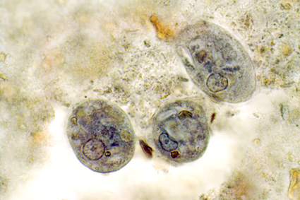
The month of September saw the publication of the first data on Blastocystis subtypes going out from Qatar. Abu-Madi and colleagues--who have already been quite prolific in terms of surveying intestinal parasitic infections in Qatar--studied the positive rate of Blastocystis in 608 apparently healthy subjects arriving in Qatar for the first time, identifying a prevalence of 71% as identified by PCR. Strikingly, the positive rate by microscopy of the corresponding samples was only 7%. Three subtypes were idenfied, with ST3 being the most common subtype, followed in prevalence by ST1 and ST2. The study is important for at least two reasons: It confirms the drawback of basing Blastocystis epidemiological research on data generated using microscopy alone, and it confirms the virtual absence of ST4 outside of Europe.

Increased sensitivity of PCR relative to microscopy was also confirmed in a study carried out in Malaysia (I presume) by Ragavan and colleagues. This group surveyed the Blastocystis positivity rate among IBS and non-IBS patients analyzing colonic aspirates, including a total of 109 individuals. Given the data available on Blastocystis prevalence, I was quite surprised to learn that this group failed to detect Blastocystis in any of the samples by microscopy and culture. Using PCR (the subtype-specific [STS] primers were used as diagnostic primers), the group identified Blastocystis in 6 IBS patients and 4 non-IBS patients. Also these figures appear quite low. However, there is very little information available on the non-IBS patients, and since all study individuals were subject to colonscopy, this group of individuals might be suffering chronic and potentially severe intestinal disease, including for instance colorectal cancer, inflammatory bowel disease, etc., which would explain the low prevalence of Blastocystis observed among these individuals. Indeed, evidence is accumulating that the more "gut healthy" you are, the larger the probability of being Blastocystis-positive. I noticed that the colonic aspirates were spun down using 3,000 rpm prior to culture and microscopy; this process might have had an impact on cell viability and morphology; still, DNA should be detectable following this process. Meanwhile, we recently showed (Scanlan et al., 2015) that the sensitivity of the STS primers is relatively low, which is why the use of real-time PCR is recommendable for PCR-based screening. To see an example of how the STS primers perform relative to barcoding primers, go here (Suppl Table 2).
Moreover, care should be taken when reading this paper, since I'm fairly convinced that the subtype terminology used in the study is different from the consensus terminology (Stensvold et al., 2007). It says that the subtypes detected included ST2, ST3, ST4, and ST5; if this reflects the terminology that went along with the original description of the STS primers, these subtypes correspond to ST7, ST3, ST6, and ST2, which to me would be a more likely subtype distribution, taking this particular region into consideration, and given the fact that ST5 appears to be extremely rare in humans. 

It's always interesting to expand on the natural host spectrum of Blastocystis. The parasite has been found in a perplexing array of hosts, but some host specificity has been observed. When it comes to animals held by humans as livestock or pets, we know that pigs and cattle are commonly, if not consistently, colonised by Blastocystis with some quite specific subtypes. With regard to pets, dogs and cats have been found positive, but there seems to be increasing evidence that these animals are not natural hosts (see also Wang et al., 2013). Osman and colleagues, recently published a survey on Cryptosporidium and Blastocystis in dogs using sensitive molecular methods, demonstrating a prevalence of Blastocystis of only about 3%. Moreover, the subtypes 2 and 10 were found, and ST10 is found mostly in cattle, and never before in dogs, as far as I know, which could suggest accidental colonisation - and possibly not a very long-lasting one. Similarly, when humans are found to be colonised with subtypes rarely found in humans, such as ST6, ST7, and ST8, it would be interesting to know for how long these subtypes are capable of "staying put" in the human intestine.

References

Abu-Madi M, Aly M, Behnke JM, Clark CG, & Balkhy H (2015). The distribution of Blastocystis subtypes in isolates from Qatar. Parasites & Vectors, 8 PMID: 26384209

Osman M, Bories J, El Safadi D, Poirel MT, Gantois N, Benamrouz-Vanneste S, Delhaes L, Hugonnard M, Certad G, Zenner L, & Viscogliosi E (2015). Prevalence and genetic diversity of the intestinal parasites Blastocystis sp. and Cryptosporidium spp. in household dogs in France and evaluation of zoonotic transmission risk. Veterinary Parasitology PMID: 26395822   

Ragavan, N., Kumar, S., Chye, T., Mahadeva, S., & Shiaw-Hooi, H. (2015). Blastocystis sp. in Irritable Bowel Syndrome (IBS) - Detection in Stool Aspirates during Colonoscopy PLOS ONE, 10 (9) DOI: 10.1371/journal.pone.0121173  

Scanlan PD, Stensvold CR, & Cotter PD (2015). Development and Application of a Blastocystis Subtype-Specific PCR Assay Reveals that Mixed-Subtype Infections Are Common in a Healthy Human Population. Applied and Environmental Microbiology, 81 (12), 4071-6 PMID: 25841010   

Stensvold CR, Suresh GK, Tan KS, Thompson RC, Traub RJ, Viscogliosi E, Yoshikawa H, & Clark CG (2007). Terminology for Blastocystis subtypes--a consensus. Trends in Parasitology, 23 (3), 93-6 PMID: 17241816

Wang W, Cuttell L, Bielefeldt-Ohmann H, Inpankaew T, Owen H, & Traub RJ (2013). Diversity of Blastocystis subtypes in dogs in different geographical settings. Parasites & vectors, 6 PMID: 23883734

Thursday, May 1, 2014

This Month In Blastocystis Research (APR 2014)

Due to all sorts of activities I have not been able to update myself with 'novelties' in the scientific Blastocystis literature lately.

Instead, I would like to highlight two review/opinion papers on the use of PCR-based methods for diagnosis of intestinal parasitic infections in the clinical microbiology laboratory.

Both papers have been published very recently (actually one is still 'in press'). The first is co-authored by Jaco J Verweij and myself, and appears in the April issue of 'Clinical Microbiology Reviews'. This paper aims to provide a relatively systematic review of the extent and relevance of PCR- and sequencing-based methods for diagnosis and epidemiology studies of intestinal parasites, and is as such an inventory of all sorts of DNA-based diagnostic and typing modalities for individual protists and helminths.

The second one is authored solely by Jaco J Verweij and is currently in the 'first online' section in the journal 'Parasitology'. This paper offers a discussion of the application of PCR-based method as a supplementary tool or a substitute for conventional methods (microscopy, antigen detection, etc.). Dr Verweij deals with central questions such as 'Is Molecular Detection Good Enough?' and 'Is Molecular Detection Too Good To Be True?'.

And so these two papers complement each other quite well. For those interested in the very low prevalence of intestinal helminth infections in the Western world, the latter paper has a table which summarizes some quite stunning data.

Although DNA-based methods currently in use do have quite a few limitations, I do believe that for a long while the application of species- and genus-specific PCR methods (real-time PCR, conventional PCR + sequencing, etc.) will appear relevant and state-of-the-art. Dr Verweij, I and a few of our colleagues around the world are currently discussing to which extent next generation sequencing methods can be used to
  • generate data that can assist us in identifying the role of pro- and eukaryote microbial communities in health and disease
  • serve as a tool to generate sequences that can be processed by designated software and thereby identify patterns of microbial communities associated with various disease and health conditions
To this end, at the Laboratory of Parasitology, Statens Serum Institut, we are currently assisting in the development of a software called BIONmeta. BION meta is an open-source package for rRNA based pro- and eukaryote community analysis. Like Qiime and Mothur it is open source but with a growing number of advantages. The package has so far been developed mostly by Niels Larsen (DK), one of the original Ribosomal Database Project authors. It is as yet unpublished, but has been selected for in-house trial-use by companies and institutions that also partly sponsor its development.When relevant, I'll post more information on this software.

References:

Verweij JJ, & Stensvold CR (2014). Molecular testing for clinical diagnosis and epidemiological investigations of intestinal parasitic infections. Clinical Microbiology Reviews, 27 (2), 371-418 PMID: 24696439

Verweij, JJ. (2014). Application of PCR-based methods for diagnosis of intestinal parasitic infections in the clinical laboratory Parasitology, 1-10 DOI: 10.1017/S0031182014000419

Wednesday, September 4, 2013

Yes, we do take orders!

I get an increasing amount of requests for Blastocystis testing (and testing for other parasites as well, for instance Dientamoeba fragilis). Initially, I was happy to do this for free, but now the requests are so regular that I need to add a fee to the tests.

And yes, we do take orders! As the regular reader of this blog would know by now, I run the part of our  Parasitology lab at Statens Serum Institut, Copenhagen, that deals with Blastocystis diagnostics and diagnostics for intestinal parasites in general. I have been developing and optimising molecular Blastocystis diagnostics for years, something which is also witnessed by my scientific production. Please note that we take orders only from health authorities. This means that if you want to have samples tested in our lab, you should contact your GP/specialist/whatever, and have him/her put the order through.

For general screening, I recommend real-time PCR analysis. For evaluation of treatment I recommend adding Blastocystis culture (a positive culture means ongoing Blastocystis infection, while DNA-based tools such as our real-time PCR will detect both dead and live organisms). We also perform subtyping of Blastocystis upon request.

In cases where colleagues want to outsource diagnostic work related to research, we are currently opening up for the possibility of testing large panels of faecal samples (fresh, frozen, or ethanol-preserved) for Blastocystis, Dientamoeba fragilis or other parasites by molecular assays (including DNA extraction) - and - if requested - in combination with traditional microscopy of faecal concentrates.

A selection of our analyses for parasites can be viewed here.

Our parasitology lab is merged with the mycology lab, and therefore we have plenty of opportunity to test the same stool sample for parasites and yeasts (e.g. Candida), if requested. As a new feature, Blastocystis+Dientamoeba+Candida analyses can now be requested in combination as a 'package' with a discount. We are happy to send out test tubes and transport envelopes, but I repeat that charges will apply.

Research-wise, we are currently taking different approaches to detecting and differentiating non-human eukaryotic DNA/RNA in human faecal samples, among these the GUT 18S approach.

For further inquiries and information, please do not hesitate to contact me (contact details can be found here).

Relevant articles on molecular diagnostics for Blastocystis detection and subtyping:

Stensvold CR, Ahmed UN, Andersen LO, & Nielsen HV (2012). Development and evaluation of a genus-specific, probe-based, internal-process-controlled real-time PCR assay for sensitive and specific detection of Blastocystis spp. Journal of Clinical Microbiology, 50 (6), 1847-51 PMID: 22422846

Stensvold CR (2013). Comparison of sequencing (barcode region) and sequence-tagged-site PCR for Blastocystis subtyping. Journal of Clinical Microbiology, 51 (1), 190-4 PMID: 23115257

Friday, May 10, 2013

Cell Symposium: Microbiome & Host Health - Lisbon 2013

My colleagues from Statens Serum Institut and I are heading to Lisbon, Portugal, tomorrow morning to attend the Cell Symposium on Microbiome and Host Health (link may be really busy now).

We are bringing a poster displaying some of our work related to our GUT18S project: A Novel Approach For Eukaryotic Phylogenetic Interrogation Of Clinical Samples Using Next Generation Sequencing Of SSU rRNA Genes; a pdf version of the poster can be downloaded here.

The GUT18S work is partly funded by the Marie Curie Actions (FP7) program.

Thursday, May 9, 2013

YouTube Video on Blastocystis Subtyping

For those who want to venture into Blastocystis subtyping - the easy way - I've recorded and uploaded a video on YouTube fyi.


For even more information, please visit a selection of relevant blog posts here.

Monday, April 29, 2013

'Invasive Blastocystis' in ECCMID 2013

ECCMID - the annual European Congress of Clinical Microbiology and Infectious Diseases (hosted by ESCMID) is currently taking place in Berlin. This year, I'm not attending, but I've been scanning the abstract book for 'Blastocystis', and it appears that an oral presentation was scheduled for yesterday in the "Emerging Infectious Diseases" section:

First of all: it's great to see fellow researchers screening larger (i.e. hundreds) of faecal DNAs by PCR for Blastocystis. I wish more people would do that to produce reliable data on prevalence and subtypes.

Now, as I've already mentioned, there are currently mainly two methods in use for subtyping, barcoding and STS PCR, and recently I evaluated these. To cut a long story short, barcoding is recommended for subtyping, since the STS method, which was used in the study by Tarasova et al. (abstract), appears to miss the majority of ST4 strains (the major genotype), and moreover, no STS primers exist for ST8 and ST9 (or any of the other 8 subtypes identified to date, but which have only been found in animals). So, the subtype data found in this study should be interpreted with this in mind.

Importantly however, I'm not sure whether the authors used the original Yoshikawa STS terminology or the terminology acknowledged in our 2007 consensus.

First, let us assume that consensus terminology is used. Then it's surprising to find ST5 in human samples in the first place, and finding a ST5 prevalence of 45% in a cohort of humans included in a larger study like this is very unlikely based on current evidence of more than 3,000 observations from all over the world, where the overall prevalence of ST5 in humans is <1%. Also, finding so much ST6 is also really striking. Also, if the consensus terminology is used, then I'm a bit puzzled why the authors put emphasis on ST7 not being found, since ST7 is relatively rare in humans.

And so let us assume that consensus terminology was not used, and the original Yoshikawa terminology was used instead. This would translate into STs 4, 6, and 7 not being detected in the CVH group. Which makes sense, since ST6 is extremely rare (at least in Europe), ST7 is only seen on occasion, and, as I said, the majority of ST4 infections are likely to go undetected by the STS method. However, ST4 appears quite common in Europe, and I suspect that it should be quite common in St Petersburg as well. But then there is one thing that comes to my mind: If ST4 infections are common, then there should be a relatively large number of samples detected by PCR which were untypable by PCR...and there is no information on untypable positive samples in the abstract...
But what is more:  STS subtype 5 translates into ST2 in consensus terminology, and similarly STS subtype 6 equals ST5 (yes, it may seem confusing, but we have provided a table in the 2007 consensus paper to make this easy). This means that no matter which of the two terminologies were used, ST5 is seen in abundance in patients with CVH in St Petersburg! Which is a very remarkable observation, and maybe more interesting than the rest of the data, which  I, by the way, find a bit difficult to follow (I expected to learn something about Blastocystis invasion, when I read the title of the abstract, but there is no data or information on invasiveness... and I'm very curious as to how the authors managed to obtain such a high number of samples from 'healthy people'! To evaluate the prevalence of Blastocystis in the control group, demographic data are needed, and a prevalence as low as 5.3% among healthy individuals makes me suspect that this control group consisted of newborns/toddlers who generally have a low prevalence of Blastocystis). Also, since when was ST1 'zoonotic'?

Anyway, often conference abstract are previews of upcoming articles, and so I expect that there will be a paper out soon from this group, and hopefully these issues will be clarified. The occasional confusion in Blastocystis epidemiology could be reduced to a minimum if everyone got into using barcoding and the Blastocystis 18S subtyping site (and go here for a video introduction to Blastocystis subtyping).

Are some citizens of St Petersburg infected by Blastocystis sp. ST5, a subtype seen primarily in livestock and African apes? Source

References:
Tarasova E, Suvorova M, Sigidaev A, Suvorov A. Blastocystis invasion in patients with chronic viral hepatitis in Saint Petersburg. ECCMID 2013 abstract O338.

Alfellani MA, Stensvold CR, Vidal-Lapiedra A, Onuoha ES, Fagbenro-Beyioku AF, & Clark CG (2013). Variable geographic distribution of Blastocystis subtypes and its potential implications. Acta Tropica, 126 (1), 11-8 PMID: 23290980

Stensvold CR (2013). Comparison of sequencing (barcode region) and sequence-tagged-site PCR for Blastocystis subtyping. Journal of Clinical Microbiology, 51 (1), 190-4 PMID: 23115257

Stensvold CR, Suresh GK, Tan KS, Thompson RC, Traub RJ, Viscogliosi E, Yoshikawa H, & Clark CG (2007). Terminology for Blastocystis subtypes--a consensus. Trends in Parasitology, 23 (3), 93-6 PMID: 17241816

Saturday, February 2, 2013

Blog Feedback

I'm very thankful for all the positive feedback I get from readers across the globe, mostly by email. Due to time limits I can only respond to 5-10% of the mail, and I'm sorry for not getting back to the rest of you.

Meanwhile, this blog currently holds more than 60 posts, and you will also find a lot of key words in the right side bar, so take your time and browse a few posts or look up a few relevant key words, -  you might find an answer to one or more  of your questions.

Having said that, I try to read all my email, and I am listening! The feedback and questions that I get are vital for our work and help us identify the avenues that we need to take to unveil the many mysteries of Blastocystis.

And let me just say this for now: A proper microbiological work-up (by state-of-the-art methods, including PCR for intestinal parasites), is something that is offered on a routine basis in only very few laboratories, and also the number of clinically orientated Blastocystis research centres can be counted on one hand, I believe. Subtyping of Blastocystis is currently done mostly in epidemiological surveys (as part of research projects), and I suspect that our lab is one of the very few labs in the world doing subtyping on a routine basis.

Oh, and I've been asked by some readers about how to get blog updates. It's easy: You can follow this blog by email, - just scroll down and find "follow by email" in the right side bar and enter your email address. You can also subscribe to posts via atom (go to the very bottom of the page).

And then here's a little something about stomach acidity and intestinal microbiota from Scientific American, - but make sure to read the comments underneath the post too!
 

Sunday, September 2, 2012

Bugs Galore!

After spending more than 8 years in clinical microbiology with special reference to parasitology, I’ve come to realise that it truly is a bug’s life! Use of nucleic acid-based methods such as PCR in routine clinical microbiology diagnostic labs have revealed that single-celled parasites are colonising the intestine of up to 50% of the Danish population! And so what? Well, this finding has several implications.

A couple of months ago I revisited Why it is a bugs life by Jörg Blech (The Guardian (2002)). Speaking of numbers, - I wonder which one is the most successful eukaryote in terms of numbers? Blastocystis? Dientamoeba? Or any other “Parasite sp.”? After realising that microscopy methods allow us to see only the very tip of the iceberg and after adding PCR to our routine diagnostics, we have found a few examples of “novel” parasitic species and many more may be in store for us. Morphologically identical organisms, such as those belonging to Iodamoeba bütschlii, may be found in both human and non-human hosts and may differ genetically across the nucelar small subunit rRNA gene by up to more than 30%! This is quite astonishing given the fact that the difference between human and murine small subunit rDNA is about 1%! Since these data have been established only recently, obviously no one knows the respective clinical significance of these morphologically similar but genetically very different lineages, and further studies may reveal differences in pathogenicity as seen in other amoebic genera. Blastocystis and Entamoeba coli are somewhat similar examples.

Our results reveal that faecal-oral transmission is much more common in Denmark - a highly industrialised country where drinking water comes from waterworks (i.e. no surface water supplies), where outbreaks even due to bacteria are scarce, and where authorities spend 1.2 billion DKK on food safety and control. Today, 90% of dwellings in Denmark (5.6m citizens) are connected to efficient sewage systems, and Denmark has more than 1,400 treatment plants to purify wastewater from households, businesses and institutions. But somewhere the chain pops off… Even in Denmark it is “bugs galore”, which means that faecal exposure is much more common that we would probably like to think. Intestinal protists (primarily Blastocystis and Dientamoeba) are telltales of exposure to faecal contamination and faecal-oral transmission.

In Denmark, 90% of dwellings are connected to efficient sewage systems, and the country has more than 1,400 treatment plants.

However, we might also learn to see these parasites as other types of indicators. In our experience Danish patients with inflammatory bowel disease (IBD) represent a cohort of people whose gut flora is remarkably different from that of other cohorts (patients with irritable bowel syndrome (IBS) and patients with non-IBD/non-IBS diarrhoea): Apparently IBD patients don’t harbour parasites. This can in part be explained by the fact that some IBD patients have had bowel resection, but even IBD patients with in intact bowel system are generally negative for parasites.

We know that in highly developed countries the prevalence of helminth infections has gone down over the past few decades due to improved hygiene measures, but maybe also due to other reasons, which have not been clarified, but as we have seen, many of us are still positive for one or more intestinal parasites. However, most IBD patients do not have any parasites at all. This correlates well with the hygiene hypothesis, and it may be so that not only helminths, but also amoebae, which are able to colonise our guts for months and even years, may be co-responsible for 1) preventing us from developing inflammatory bowel disease and other autoimmune diseases by immunomodulatory mechanisms, and 2) maintaining a sound intestinal flora and ecology. Or is it so that these protists are dependent on a certain gut ecology or gut flora in order to colonise our intestines for a longer period, and in this way, they can be seen as indicators of a certain gut microbiota? Do they have any modulatory functions or do they happen to "lead their own life"?

As a parasitologist and worshipper of most things eukaryotic, I was both pleased and disconcerted after leaving the MetaHIT conference in Paris in March. Pleased, since the stratification of people into enterotypes and correlation of enterotypes to disease phenotypes suited my naïve, B/W perception of the world, but disconcerted since all presentations and posters addressed only bacteria (and virus to a minor extent, - maybe one on archaea even?). But, how about intestinal yeasts and parasites? Where in the gene catalogues and pools of metagenomic data could I find information on eukaryotes? Nowhere. Which hopefully boils down to methodological limitations rather than absence of interest.

The concept of paving an avenue of new knowledge with metagenomics data is holistic in its approach, but it currently fails to encompass a common part of the intestinal microbiota, possibly due to methodological limitations. However, we are probably facing the imminent inclusion of eukaryotic data in metagenomic studies, and this will enable us to investigate the potential role of intestinal protists and maybe yeasts as biomarkers of certain enterotypes and maybe even disease or health phenotypes.

Further reading:

Stensvold CR, Lebbad M, & Clark CG (2012). Last of the human protists: the phylogeny and genetic diversity of Iodamoeba. Molecular biology and evolution, 29 (1), 39-42 PMID: 21940643

Stensvold CR (2012). Thinking Blastocystis out of the box. Trends in parasitology, 28 (8) PMID: 22704911

Friday, August 10, 2012

Is This A New Subtype?

To quote one of my colleagues attending the recent IWOP 2012 meeting in Tarrytown, NY, Blastocystis subtyping in humans and animals is becoming 'trendy', and so we keep trying to advocate for a standardisation of the metholodology of Blastocystis subtyping.

We recently changed the title of our page at www.pubmlst.org/blastocystis so that now it is called Blastocystis Subtype (18S) and Sequence Typing (MLST) Databases, and we added some text to front page:

In terms of genetic markers, the barcode region (Scicluna et al., 2006) is by far the best represented in publicly available sequence databases, and the correct subtype can be identified by BLAST analysis in the sequence database at the present site. Blasting against this database has the added advantages, compared to using GenBank, of automatically assigning allele types to the SSU-rDNA as well as using the consensus subtype nomenclature (unlike GenBank where the subtype is included only if one was part of the accession submission and no attempt to impose a standard nomenclature is made). In case the sequence does not match any of the ones in the database despite full coverage of the region, this indicates that the sequence represents a new allele or maybe even a new subtype depending on the amount of variation. If a new subtype is suspected, we suggest doing PCR and sequencing of the complete SSU rRNA gene with subsequent phylogenetic analysis using reference sequences.

Now, the last bit is extremely important. We have seen examples of researchers (including ourselves!) assigning sequences to a new a subtype in the absence of complete SSU rDNA data (in fact complete sequences for ST10-ST14 are not yet publicly available!). Doing so has a least two major limitations/drawbacks: Far from all SSU rDNA regions have been validated as being representative of the whole SSU rRNA gene in terms of phylogenetic analysis, and therefore phylogenetic inferences based on non-validated regions may have little or at least less support than anticipated. Moreover, if someone analyses e.g. position 600-1600, and phylogenetic analysis based on this region reveals a potentially new subtype, this makes it impossible for his/her colleague who has data covering positions 1-600 from a Blastocystis isolate that may also represent a new subtype to ascertain whether it might be same subtype (see example below)!

Obtaining complete SSU rDNA sequences directly from faecal DNA may be a cumbersome task but is sometimes possible by combining sequence-specific primers with low-specificity primers such as the RD5 and the RD3 primers (Clark, 1997). If a cultured isolate is available, obviously this makes complete SSU rDNA sequencing much easier.

While it appears that the number of subtypes occurring in humans stays around 9, our gut feeling is that we are yet to uncover quite a few subtypes colonising non-human mammals, and it's great to see an increasing number of teams exploring the genetic diversity of Blastocystis. For instance, Dr Ronald Fayer and his group recently published exciting data on a new Blastocystis subtype in cattle, which they named ST14 (Fayer et al., 2012).

Importantly, caution should be taken to avoid creating confusion in subtype terminology. Confusion can arise when independent researchers assign the same new subtype name (e.g. ST14, ST15, etc.) to novel sequences which in fact belong to different ribosomal lineages, or when incomplete SSU rDNA sequence data are used; this situation was seen recently, when Petrasova et al. (2011), assigned a Colobus sequence to ST5, although it was in fact a ST13 sequence (Clark et al., in press); the situation arose, since Petrasova et al. (2011) did not have data covering the region currently available for ST13 (Parkar et al., 2010), and therefore believed that their sequence was a unique ST5 variant. As for ST14, less than 500 bp are currently available, and these 500 bp are not in the barcode region, making it difficult for all teams using barcoding to compare their data. And so we would like to advocate for making complete SSU rDNA sequences publicly available (Genbank) for potentially new subtypes, for at least two reasons:

1. Phylogenetic inferences based on the complete SSU rDNA will be more robust than those obtained from analysing shorter sequence streches.

2. Complete seqeunces are needed for reference since subtype screening typically includes a single round PCR such as barcoding (Scicluna et al., 2006) amplifying about 550 bp; in the situation where complete SSU rDNAs are available for all known subtypes, it will be quick to analyse, whether a sequence may represent a new subtype, since this will be independent on the SSU rDNA region studied.We therefore hope that complete SSU rDNA sequences will soon be made publicly available for ST10-ST14.

So, when does a complete SSU rDNA sequence represent a new subtype? Well, we have a review paper in press in Advances in Parasitology on recent developments in Blastocystis research, which will be published in less than six months probably, and which also touches on this topic; once the paper is published, I will try and make a summary our thoughts on this...

Further reading:


Clark CG (1997). Extensive genetic diversity in Blastocystis hominis. Molecular and biochemical parasitology, 87 (1), 79-83 PMID: 9233675

Fayer R, Santin M, & Macarisin D (2012). Detection of concurrent infection of dairy cattle with Blastocystis, Cryptosporidium, Giardia, and Enterocytozoon by molecular and microscopic methods. Parasitology research PMID: 22710524

Parkar U, Traub RJ, Vitali S, Elliot A, Levecke B, Robertson I, Geurden T, Steele J, Drake B, & Thompson RC (2010). Molecular characterization of Blastocystis isolates from zoo animals and their animal-keepers. Veterinary parasitology, 169 (1-2), 8-17 PMID: 20089360

Petrášová J, Uzlíková M, Kostka M, Petrželková KJ, Huffman MA, & Modrý D (2011). Diversity and host specificity of Blastocystis in syntopic primates on Rubondo Island, Tanzania. International journal for parasitology, 41 (11), 1113-20 PMID: 21854778
 
Scicluna SM, Tawari B, & Clark CG (2006). DNA barcoding of blastocystis. Protist, 157 (1), 77-85 PMID: 16431158

Sunday, July 1, 2012

Do I Get Diagnosed Correctly?

I can tell especially from Facebook discussions that people across the globe wanting to know about their "Blastocystis status" are worried that they are receiving false-negative results from their stool tests, and that many Blastocystis infections go unnoticed. And I think I should maybe try and say a few things on this (please also see a recent blog post on diagnosis, - you'll find it here). I might try and simplify things a bit in order not to make the post too long.

Below, you'll find a tentative representation of the life cycle of Blastocystis. It is taken from CDC, from the otherwise quite useful website DPDx - Laboratory Identification of Parasites of Public Health Concern.

Proposed life cycle of Blastocystis.
 
I don't know how useful it is, but what's important here is the fact that we accidentally ingest cysts of Blastocystis, and we shed cysts that can be passed on to other hosts. The cyst stage is the transmissible stage, and the way the parasite can survive outside the body; we don't know for how long cysts can survive and remain infective. In our intestine and triggered by various stimuli, the cysts excyst, transiting to the non-cyst form, which could be called the trophozoite / "troph" stage, or to use a Blastocystis-specific term, the "vacuolar stage" (many stages have been described for Blastocystis, but I might want to save that for later!). This is possibly the stage in the life cycle where the parasite settles, thrives, multiplies, etc. You can see a picture of vacuolar stages in this blog post. Many protozoa follow this simple life cycle pattern, among them Giardia and most species of Entamoeba. If the stool is diarrhoeic and you are infected by any one or more of these parasites, it may be so that only trophozoites, and, importantly, no cysts, are shed! This has something to do with reduced intestinal transit time and, maybe more importantly, the failure of the colon to resorb water from the stool which means that the trophozoites do not get the usual encystation stimuli. Importantly, trophozoites are in general non-infectious.

There is documentation that once colonised with Blastocystis, you may well carry it with you for years on end, and as already mentioned a couple of times, no single drug or no particular diet appears to be capable of eradicating Blastocystis - this is supported by the notion that Blastocystis prevalence seems to be increasing by age, although spontaneous resolution may not be uncommon, - we don't know much about this. Now, although day-to-day variation in the shedding of Blastocystis has been described, it is my general impression that colonised individuals may shed the parasite with each stool passage, and well-trained lab technicians/parasitologists will be able to pick up Blastocystis in a direct smear (both cysts and trophs may be seen). To do a direct smear you simply just mix a very small portion of the stool with saline or PBS on a slide, put a cover slip over it and do conventional light microscopy at x200 (screening) or x400 (verification). Very light infections may be difficult to detect this way, and if you don't have all the time in the world, a direct smear may not be the first choice.

The "king" of parasitological methods, however, is microscopy of faecal concentrates (Formol Ethyl Acetate Concentration Technique and any variant thereof), which is remarkable in its ability to detect a huge variety of parasites. Especially cysts of protozoa (e.g. Giardia and Entamoeba) and eggs of helminths (e.g. tapeworm, whipworm and roundworm) concentrate well and are identified to genus and species levels based on morphology. The method is not as sensitive as DNA-based methods such as PCR, but as I said, has the advantage of picking up a multitude of parasites and therefore good for screening; PCR methods are targeted towards particular species (types) of parasites. A drawback of the concentration method is that it doesn't allow you to detect trophzoites (i.e. the fragile, non-cystic stage), and, as mentioned, diarrhoeic samples may contain only trophozoites and no cysts...

In many countries it is very common for people to be infected by both protozoa and helminths, and in those countries microscopy of faecal concentrates is a relevant diagnostic choice. In Denmark and many Western European countries, the level of parasitism is higher than might be expected (from a hygiene and food safety point of view) but due to only few parasitic species. Paradoxically, the intestinal parasites that people harbour in this part of the world are parasites that do not concentrate well. They are mainly:

1) Blastocystis
2) Dientamoeba fragilis
3) Pinworm (Enterobius vermicularis)

Only troph stages have been described for Dientamoeba fragilis and it may be transmitted by a vector, such as pinworm (look up paper by Röser et al. in the list below for more information); this mode of transmission is not unprecedented (e.g. Histomonas transmission by Heterakis). Eggs of pinworm may be present in faeces, but a more sensitive method is the tape test.

Now, Blastocystis often disintegrates in the faecal concentration process, and while you might be lucky to pick up the parasite in a faecal concentrate, you shouldn't count on it, and hence the method is not reliable, unless the faecal sample was fixed immediatley after being voided. This is key, and also why fixatives are used for the collection of stool samples in many parts of the world - to enable the detection of fragile stages of parasites. There are many fixatives, e.g. SAF (sodium acetate-acetic formalin), PVA (poly-vinyl alcohol) and even plain formalin will do the trick if it's just a matter of preserving the parasite in the sample. If SAF or PVA is used, this allows you to do permanently stained smears of faecal concentrates, and you will be able to pick up not only cysts of protozoa, but even trophozoites. Trichrome and iron-haematoxylin staining are common methods and are sensitive but very time-consuming and may be related to some health hazards as well due to the use of toxic agents. But this way of detecting parasites is like good craftmanship - it requires a lot of expertise, but then you get to look at fascinating structures with intriguing nuclear and cytoplasmatic diagnostic hallmarks. Truly, morphological diagnosis of parasites is an art form! Notably, samples preserved in such fixatives may be useless for molecular analyses.

Iron-haematoxylin stain of trophozoites of Entamoeba coli
(note the "dirty" cytoplasm characteristic of E. coli).
Source: http://www.atlas-protozoa.com

At our lab we supplement microscopy of faecal concentrates with DNA-based detection of parasites. For some clinically significant parasites, we do a routine screen by PCR, since this is more sensitive than microscopy of faecal concentrates and because this is a semi-automated analysis that involves only DNA extraction, PCR and test result interpretation, which are all things that can be taught easily. Major drawbacks of diagnostic PCR is that you cannot really distinguish between viable (patent infection) and dead organisms (infection resolving, e.g. due to treatment). This is why, in the case of Blastocystis, you may want to do a stool culture as well (at least in post-treatment situations), since only viable cells will be able to grow, obviously.

Two diagnostic real-time PCR analyses have been published, one using CYBR Green and one using a TaqMan probe.

Now, it certainly differs from lab to lab as to which method is used for Blastocystis detection. Some labs apparently apply thresholds for number of parasites detected per visual field, and only score a sample positive if more than 5 parasites per visual field have been detected. I see no support for choosing a threshold, since 1) we do not know whether any Blastocystis-related symptoms are exacerbated by parasite intensity, 2) the number of parasites detected in a faecal concentrate may depend on so many things which have nothing to do with the observer (fluctuations in shedding for instance), and 3) the pathogenic potential of Blastocystis may very well depend on subtype.

If Blastocystis was formally acknolwedged as a pathogen, like Giardia, standardisation of methods would have happened by now. Meanwhile, we can only advocate for the use of PCR and culture if accurate diagnosis of Blastocystis is warranted, while permanent staining of fixed faecal samples constitutes a very good alternative in situations where PCR is not an option.

I have the impression that some labs do DNA-based detection of microbes, including protozoa, and that a result such as "taxonomy unknown" is not uncommon. I don't know how these labs have designed their molecular assays, and therefore I cannot comment on the diagnostic quality and relevance of those tests... it also depends on whether labs do any additional testing as well, such as the more traditional parasitological tests. However, we do know that there is a lot of organisms in our intestine, for which no data are available in GenBank, which is why it is sometimes impossible to assign a name to e.g. non-human eukaryotic DNA amplified from a stool sample.

* More than 1 billion people may harbour Blastocystis.
* Blastocystis is found mainly in the large intestine.
* 95% of humans colonised by Blastocystis have one of the following subtypes: ST1, ST2, ST3, ST4.
* DNA-based detection combined with culture ensures accurate detection of Blastocystis in stool samples and enables subtyping and viability assessment.


Further reading:

Poirier P, Wawrzyniak I, Albert A, El Alaoui H, Delbac F, & Livrelli V (2011). Development and evaluation of a real-time PCR assay for detection and quantification of blastocystis parasites in human stool samples: prospective study of patients with hematological malignancies. Journal of clinical microbiology, 49 (3), 975-83 PMID: 21177897

Röser D, Nejsum P, Carlsgart AJ, Nielsen HV, & Stensvold CR (2013). DNA of Dientamoeba fragilis detected within surface-sterilized eggs of Enterobius vermicularis. Experimental parasitology, 133 (1), 57-61 PMID: 23116599

Scanlan PD, & Marchesi JR (2008). Micro-eukaryotic diversity of the human distal gut microbiota: qualitative assessment using culture-dependent and -independent analysis of faeces. The ISME journal, 2 (12), 1183-93 PMID: 18670396

Stensvold CR, Ahmed UN, Andersen LO, & Nielsen HV (2012). Development and Evaluation of a Genus-Specific, Probe-Based, Internal-Process-Controlled Real-Time PCR Assay for Sensitive and Specific Detection of Blastocystis spp. Journal of clinical microbiology, 50 (6), 1847-51 PMID: 22422846

Stensvold CR, Arendrup MC, Jespersgaard C, Mølbak K, & Nielsen HV (2007). Detecting Blastocystis using parasitologic and DNA-based methods: a comparative study. Diagnostic microbiology and infectious disease, 59 (3), 303-7 PMID: 17913433

Stensvold CR, & Nielsen HV (2012). Comparison of microscopy and PCR for detection of intestinal parasites in Danish patients supports an incentive for molecular screening platforms. Journal of clinical microbiology, 50 (2), 540-1 PMID: 22090410