Showing posts with label molecular epidemiology. Show all posts

Monday, July 29, 2013

Birds of America

Yesterday evening, I was watching another compelling BBC production, broadcast on Danish television: Earthflight, North America. In quite a unique way, the viewers got the rare opportunity to see through the eyes of birds such as eagles, geese, and pelicans and follow birds as they were migrating, escaping, hunting for prey, etc. It made me think of the 19th century masterpiece 'Birds of America' by John James Audubon, which can be viewed in the National History Museum in London. The book features 435 stunning hand-coloured plates that show birds life-size, in natural positions and in their natural habitat.

One of the things that I find interesting - and quite unexplored - is Blastocystis in birds. By 'unexplored' I mean that relatively little sampling has been done, and so the number of observations of Blastocystis in birds is still limited compared to other types of hosts. However, there is a brand new paper out in 'Infection, Genetics and Evolution' which includes observations on Blastocystis in birds (of America!).

You see, I was invited in on a study by colleagues in Colombia who had access to DNA from quite a few faecal samples from a number of host species, including feral birds, and what we found confirms the quite unambiguous trend seen so far: Birds - no matter where on this planet - appear to be colonised mainly by ST6 and ST7. As a matter of fact, in the present study only ST6 was seen in almost 50 Colombian passerine birds of varying species, most of which I believe are limited in geographical distribution to the Americas: Passer domesticus, Thraupis episcopus, Oryzoborus maximiliani, Sicalis flaveola, and Petrochelidon pyrrhonota. Moreover, only one allele of ST6, allele 122, was identified. Notably, the prevalence of Blastocystis in the sampled bird population was 90%. I believe that this is the first official report on Blastocystis in passeriformes. Other major groups of birds previously sampled include galliformes, anseriformes, and ratites (Stensvold et al., 2009; Alfellani et al., 2013).

Other subtypes have been reported in birds (Alfellani et al., 2013), but due to the very low number of samplings these subtypes may be more or less co-incidental/abberant findings. Of note, some samples from birds have been untypable. I have a slight recollection of detecting ST3 in Icelandic rock ptarmigans (in mixed ST infection) collected by Dr Karl Skírnission, but that certainly needs confirmation.

Bird contact/bird droppings - a significant source of Blastocystis in humans? Me feeding some 'Birds of Australia'. Photo by Dr Rebecca J Traub.

ST6 is very rarely seen in humans in Europe. In other parts of the world, for instance in Egypt and some Asian countries, ST6 appears relatively common, but we do not know much about 'bird subtypes' in those particular regions. Also, the situation in the US and Canada is more or less completely unknown (Blastocystis subtyping is something that appears not to attract research groups in North America apart from the one led by Dr Ron Fayer in Beltsville, Maryland).

ST7 is occasionally seen in humans in countries such as Sweden and Denmark. But in my - still limited - experience, individuals infected by these subtypes are not necessarily prone to 'suffer more' from intestinal symptoms than those who do not have these subtypes. While human cases of ST6 (and ST7) may represent cases of zoonotic transmission, it is far to early to draw any conclusions on this. It would be important to compare ST6 and ST7 18S alleles from humans and birds. MLST typing systems for these two subtypes are not yet available, but 18S analysis in itself may prove valuable for molecular epidemiological analyses as in the case of other subtypes (Stensvold et al., 2012).

Walton Ford: "Falling Bough" (Source). You will also see the now extinct Passenger Pigeon in 'Birds of America'.

References:

Ramírez JD, Sánchez LV, Bautista DC, Corredor AF, Flórez AC, & Stensvold CR (2013). Blastocystis subtypes detected in humans and animals from Colombia. Infection, Genetics and Evolution: Journal of Molecular Epidemiology and Evolutionary Genetics in Infectious Diseases PMID: 23886615

Alfellani MA, Taner-Mulla D, Jacob AS, Imeede CA, Yoshikawa H, Stensvold CR, & Clark CG (2013). Genetic diversity of Blastocystis in livestock and zoo animals. Protist, 164 (4), 497-509 PMID: 23770574

Stensvold CR, Alfellani M, & Clark CG (2012). Levels of genetic diversity vary dramatically between Blastocystis subtypes. Infection, Genetics and Evolution: Journal of Molecular Epidemiology and Evolutionary Genetics in Infectious Diseases, 12 (2), 263-73 PMID: 22116021

Stensvold CR, Alfellani MA, Nørskov-Lauritsen S, Prip K, Victory EL, Maddox C, Nielsen HV, & Clark CG (2009). Subtype distribution of Blastocystis isolates from synanthropic and zoo animals and identification of a new subtype. International Journal for Parasitology, 39 (4), 473-9 PMID: 18755193

Wednesday, July 10, 2013

This Month In Blastocystis Research (JUL 2013)

The open access journal 'Tropical Parasitology' (published by the Indian Academy of Tropical Parasitology) has included a symposium on Blastocystis in their January-June (Vol. 3) issue (available here). The symposium comprises three papers; one is on "taxonomy, biology and virulence", the next is on genetic diversity and molecular methods for diagnosis and epidemiology, and the last one is on treatment controversies. I believe that it may take quite a while before these papers will appear in PubMed.

The first paper written by Drs Parija and Jeremiah sums up a few of the aspects related to (especially historical) taxonomic issues and very little on the actual biology of Blastocystis. Meanwhile, there is quite a substantial section on Blastocystis morphology. Regarding virulence, the authors mention the possibility that differences in virulence may be due to differences in subtypes, but that subtyping alone does not predict pathogenicity which in part may be due to varying levels of intra-subtype genetic variation. The authors also briefly mention some of the morphological and phenotypical observations that have been associated with 'pathogenic Blastocystis', such as the amoeboid stage, large cells, rough surface, slow growth rate, and increased binding to lectins. It is always interesting to speculate on such associations, but it must be kept in mind that results from in-vitro experiments may not necessarily reflect in-vivo situations.

One topic that keeps popping up in the literature - and also in two of the papers here in this symposium - is the possibility of 'amoebic forms' of Blastocystis being associated with symptomatic infection. This hypothesis was introduced in 2006 by Tan and Suresh, I believe; Scanlan (2013) speculated that amoeboid forms might be the nutrient acquiring form potentially selecting for bacterial virulence or certain bacterial communities through grazing; please go here for more thoughts from a previous blog post.

My own experience on Blastocystis morphology mainly stems from looking at cultures, and since we practically only get isolates from patients with gastrointestinal disease, I don't know what Blastocystis cultures from asymptomatic individuals look like. A dear colleague of mine - Marianne Lebbad, a brilliant Swedish parasitologist with many years in business - sent me the picture below (light microscopy of a faecal concentrate) and speculates that Blastocystis might be able to form groups/clusters of cells, maybe even with the ability to form a mono-layer on the surface of the gut mucosa? I've never observed the cluster formation in cultures, but then again, we have no idea of whether the stages seen in in vitro cultures (microaerophilic environment) are identical to the in vivo stages (strictly anaerobic), and exactly how Blastocystis lives and multiplies in the colon... Anyway, the idea of biofilm comes into mind. It would be nice to learn more from colleagues with a similar experience.

Light microscopy of Blastocystis apparently forming a cluster of cells; we wonder whether the cells are in fact 'glued' together and if so, how? Courtesy of Dr Marianne Lebbad.

Moving on to the next paper, this one was written by me and deals mostly with issues and developments within the field of diagnostics, molecular characterisation, and molecular epidemiology. The target audience comprises clinical microbiologists and those involved in Blastocystis epidemiology and genetic diversity research. Included is a table, which is basically a reproduction of the one included in the recent paper by Alfellani et al. (2013) displaying the distribution of subtypes in humans across different geographical regions. I hope that the open access feature of this paper will prompt even more researcher into Blastocystis epidemiology! At least it is currently listed on the site as 'popular'!

The third paper in the string is written by Drs Sekar and Shanthi. These authors put emphasis on the conspicuous lack of data on the metabolic processes of Blastocystis, making it difficult to establish how to best approach antibiotic intervention; we must anticipate that with more genomic and transcriptomic data analyses arriving within a foreseeable future we will soon know much more about this. They also reiterate what has been put forth by many, namely that differences in eradication may boil down to differences in drug susceptibility, which again may be due to a variety of reasons, including genetic diversity, which is extreme in Blastocystis.

According to these authors, 'therapy should be limited to patients with persistent symptoms subsequent to a complete work up for alternative etiologies'; at the present stage this appears sensible, although clinicians would probably appreciate a clearer definition of 'symptoms'!

The review goes through some of the drugs most commonly used for treating Blastocystis, including metronidazole, paromomycin and co-trimoxazole, but also includes a few data on the use of the probiotic Saccharomyces boulardii in attempts to eradicate Blastocystis. There is not very much on the mechanisms of drug action, - it's more like a summary of data coming out from different studies, including the few placebo-controlled ones.
Regarding co-trimoxazole (which is also known as 'Bactrim' or 'Septra') this drug combo is often administered to HIV-patients prophylactically against Pneumocystis. In a study of parasites in Danish HIV patients, only 6/96 patients were given co-trimoxazole (unpublished data); two of these patients had Blastocystis. Hence, one 'alternative' way of finding out about the efficacy of co-trimoxazole on Blatocystis is to test the stools from patients undergoing long-term Pneumocystis prophylaxis comparing these patients to a cohort not receiving Pneumocystis prophylaxis but otherwise similar.

I find it a bit peculiar though to go through a review on treatment data that does not at one single point mention the need for sensitive diagnostics when evaluating courses of treatment and the identification of carriers and non-carriers. Also, there are some passages which are quite difficult for me to follow, for instance p. 36, second column, bottom section.

I hope that this symposium will inspire some of our colleagues and contribute to an increased understanding of Blastocystis.

References:

SYMPOSIUM

Parija SC & Jeremiah SS (2013). Blastocystis: Taxonomy, biology and virulence Tropical Parasitology DOI: 10.4103/2229-5070.113894
 
Stensvold CR (2013). Blastocystis: Genetic diversity and molecular methods for diagnosis and epidemiology Tropical Parasitology DOI: 10.4103/2229-5070.113896  

Sekar U & Shanthi M (2013). Blastocystis: Consensus of treatment and controversies Tropical Parasitology DOI: 10.4103/2229-5070.113901

OTHER:

Scanlan PD (2012). Blastocystis: past pitfalls and future perspectives. Trends in parasitology, 28 (8), 327-34 PMID: 22738855

Stensvold CR, Nielsen SD, Badsberg JH, Engberg J, Friis-Møller N, Nielsen SS, Nielsen HV, & Friis-Møller A (2011). The prevalence and clinical significance of intestinal parasites in HIV-infected patients in Denmark. Scandinavian Journal of Infectious Diseases, 43 (2), 129-35 PMID: 20936912  

Tan TC & Suresh KG (2006). Predominance of amoeboid forms of Blastocystis hominis in isolates from symptomatic patients. Parasitology Research, 98 (3), 189-93 PMID: 16323025

Friday, June 21, 2013

This Month In Blastocystis Research (JUN 2013)

Another paper in the string of publications coming out from the PhD study by Dr Alfellani (London School of Hygiene and Tropical Medicine) has just appeared in PubMed.

Dr Alfellani and his colleagues have done a great job in analysing a multitude of samples from humans, non-human primates and animals; I have previously blogged about their observations from studies of human and non-human primates. Moreover, they have surveyed available data in order to better discuss their own findings, and the work has contributed significantly to what today is known about the host specificity, genetic diversity, phylogeography and general molecular epidemiology of Blastocystis.

Alfellani's most recent paper is published in the journal Protist, and it deals with the 'Genetic Diversity of Blastocystis in Livestock and Zoo Animals'.

It is quite a large paper which includes a lot of new information and a comprehensive (and hopefully exhaustive) table summarising Blastocystis subtype data in all relevant hosts (humans, non-human primates, other mammals and birds).

I will highlight a couple of things from the paper:

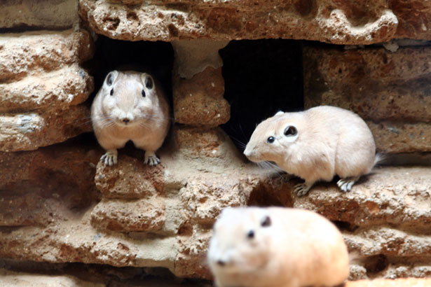
1. Apart from reporting on virtually complete SSU rDNA sequences from a couple of subtypes for which entire SSU rDNA sequences have yet not been available, we also report on three novel subtypes. Until recently, we only knew about 14 subtypes (ST1-ST14), of which ST1-ST9 can be found in humans. Now, three additional subtypes have been identified; ST15 in artiodactyls (camel and sheep) and non-human primates (chimpanzee and gibbon), ST16 in kangaroos, and ST17 in gundis.

The Gundi (Ctenodactylus gundi) is a rodent living mainly in the deserts of Northern Africa. (Source)

2. Novel data arising from analysis of faecal samples from humans and animals in Sebha, Libya, strongly indicate that humans and animals in this area are infected by different subtypes: Humans appear to carry ST1, ST2, and ST3, while synanthropic animals (artiodactyls in this case) mostly have ST5 and ST10 infections, suggesting that livestock is not a major contributor to human Blastocystis infection.

To this end, there is growing evidence of quite a substantial degree of host specificity of Blastocystis.  Even when subtypes overlap between humans and animals, we have accumulating evidence that the strains found in humans and animals are different. This means that the hypothesis that animals constitute an important reservoir of human Blastocystis infections currently has very limited support. It is my clear impression that when a strain of ST6 or ST8 is detected in humans, this strain has most probably been transmitted from an animal source. But we very rarely see these subtypes in humans, at least in Europeans.

It will be extremely interesting to see how the universe of Blastocystis subtypes unfolds... by genetically characterising strains in humans and non-human hosts, we are building up a clearer picture of transmission patterns and evolutionary biology, including our adaptation to Blastocystis, and the parasite's adaptation to us and other hosts.

It is noteworthy that we are starting to see different subtypes in rodents. We have previously thought that generally, rodents were infected by ST4. But now we know that many rodents are not infected, and we also know that rodents may harbour subtypes other than ST4.

So,17 subtypes of Blastocystis are now known. We have probably only seen the top of the iceberg, since many host species have not yet been sampled from, and it is likely that we will see quite a few STs being identified in the nearest future. To this end it is necessary to have a consensus regarding the identification of novel subtypes. Along with the Protist paper we have uploaded a supplementary file (Appendix A, TXT format) with aligned reference sequences that can be used for phylogenetic analysis,  hoping that it will be useful to our colleagues. In a future blog post I will try to address the issues of identifying new subtypes more specifically.

ST15 is one of the more interesting subtypes since it appears to have quite a low host specificity - infecting both non-human primates and artiodactyls. Yet, we have come across it only now. ST15 and ST17 are remarkable in the way that they appear to be closer related to herptile and arthropod lineages, respectively, than to lineages from mammals.

Please note that virtually complete sequences of ST10, ST13, ST14, ST15, and ST17 analysed in the study have been released in GenBank just now.

Further reading:

Alfellani MA, Taner-Mulla D, Jacob AS, Imeede CA, Yoshikawa H, Stensvold CR, & Clark CG (2013). Genetic Diversity of Blastocystis in Livestock and Zoo Animals. Protist, 164 (4), 497-509 PMID: 23770574

Alfellani MA, Stensvold CR, Vidal-Lapiedra A, Onuoha ES, Fagbenro-Beyioku AF, & Clark CG (2013). Variable geographic distribution of Blastocystis subtypes and its potential implications. Acta Tropica, 126 (1), 11-8 PMID: 23290980

Alfellani MA, Jacob AS, Perea NO, Krecek RC, Taner-Mulla D, Verweij JJ, Levecke B, Tannich E, Clark CG, & Stensvold CR (2013). Diversity and distribution of Blastocystis sp. subtypes in non-human primates. Parasitology, 140 (8), 966-71 PMID: 23561720

Thursday, May 9, 2013

YouTube Video on Blastocystis Subtyping

For those who want to venture into Blastocystis subtyping - the easy way - I've recorded and uploaded a video on YouTube fyi.


For even more information, please visit a selection of relevant blog posts here.

Monday, April 29, 2013

'Invasive Blastocystis' in ECCMID 2013

ECCMID - the annual European Congress of Clinical Microbiology and Infectious Diseases (hosted by ESCMID) is currently taking place in Berlin. This year, I'm not attending, but I've been scanning the abstract book for 'Blastocystis', and it appears that an oral presentation was scheduled for yesterday in the "Emerging Infectious Diseases" section:

First of all: it's great to see fellow researchers screening larger (i.e. hundreds) of faecal DNAs by PCR for Blastocystis. I wish more people would do that to produce reliable data on prevalence and subtypes.

Now, as I've already mentioned, there are currently mainly two methods in use for subtyping, barcoding and STS PCR, and recently I evaluated these. To cut a long story short, barcoding is recommended for subtyping, since the STS method, which was used in the study by Tarasova et al. (abstract), appears to miss the majority of ST4 strains (the major genotype), and moreover, no STS primers exist for ST8 and ST9 (or any of the other 8 subtypes identified to date, but which have only been found in animals). So, the subtype data found in this study should be interpreted with this in mind.

Importantly however, I'm not sure whether the authors used the original Yoshikawa STS terminology or the terminology acknowledged in our 2007 consensus.

First, let us assume that consensus terminology is used. Then it's surprising to find ST5 in human samples in the first place, and finding a ST5 prevalence of 45% in a cohort of humans included in a larger study like this is very unlikely based on current evidence of more than 3,000 observations from all over the world, where the overall prevalence of ST5 in humans is <1%. Also, finding so much ST6 is also really striking. Also, if the consensus terminology is used, then I'm a bit puzzled why the authors put emphasis on ST7 not being found, since ST7 is relatively rare in humans.

And so let us assume that consensus terminology was not used, and the original Yoshikawa terminology was used instead. This would translate into STs 4, 6, and 7 not being detected in the CVH group. Which makes sense, since ST6 is extremely rare (at least in Europe), ST7 is only seen on occasion, and, as I said, the majority of ST4 infections are likely to go undetected by the STS method. However, ST4 appears quite common in Europe, and I suspect that it should be quite common in St Petersburg as well. But then there is one thing that comes to my mind: If ST4 infections are common, then there should be a relatively large number of samples detected by PCR which were untypable by PCR...and there is no information on untypable positive samples in the abstract...
But what is more:  STS subtype 5 translates into ST2 in consensus terminology, and similarly STS subtype 6 equals ST5 (yes, it may seem confusing, but we have provided a table in the 2007 consensus paper to make this easy). This means that no matter which of the two terminologies were used, ST5 is seen in abundance in patients with CVH in St Petersburg! Which is a very remarkable observation, and maybe more interesting than the rest of the data, which  I, by the way, find a bit difficult to follow (I expected to learn something about Blastocystis invasion, when I read the title of the abstract, but there is no data or information on invasiveness... and I'm very curious as to how the authors managed to obtain such a high number of samples from 'healthy people'! To evaluate the prevalence of Blastocystis in the control group, demographic data are needed, and a prevalence as low as 5.3% among healthy individuals makes me suspect that this control group consisted of newborns/toddlers who generally have a low prevalence of Blastocystis). Also, since when was ST1 'zoonotic'?

Anyway, often conference abstract are previews of upcoming articles, and so I expect that there will be a paper out soon from this group, and hopefully these issues will be clarified. The occasional confusion in Blastocystis epidemiology could be reduced to a minimum if everyone got into using barcoding and the Blastocystis 18S subtyping site (and go here for a video introduction to Blastocystis subtyping).

Are some citizens of St Petersburg infected by Blastocystis sp. ST5, a subtype seen primarily in livestock and African apes? Source

References:
Tarasova E, Suvorova M, Sigidaev A, Suvorov A. Blastocystis invasion in patients with chronic viral hepatitis in Saint Petersburg. ECCMID 2013 abstract O338.

Alfellani MA, Stensvold CR, Vidal-Lapiedra A, Onuoha ES, Fagbenro-Beyioku AF, & Clark CG (2013). Variable geographic distribution of Blastocystis subtypes and its potential implications. Acta Tropica, 126 (1), 11-8 PMID: 23290980

Stensvold CR (2013). Comparison of sequencing (barcode region) and sequence-tagged-site PCR for Blastocystis subtyping. Journal of Clinical Microbiology, 51 (1), 190-4 PMID: 23115257

Stensvold CR, Suresh GK, Tan KS, Thompson RC, Traub RJ, Viscogliosi E, Yoshikawa H, & Clark CG (2007). Terminology for Blastocystis subtypes--a consensus. Trends in Parasitology, 23 (3), 93-6 PMID: 17241816

Monday, January 14, 2013

A Penny For Your Thoughts

So, what should we do about Blastocystis? What do we want to know?

I believe the imminent answer to the latter question is easy: We want to know whether it’s pathogenic, whether we should treat it and how. But I also think that there are many other interesting aspects of Blastocystis which are also of broad interest to the general public, namely: How about the many cases of asymptomatic Blastocystis carriage? What does Blastocystis do in our guts? Could it have any potentially beneficial impact on our health?

Given the fact that Blastocystis has not been implicated in any outbreaks (admittedly: I guess that no one actually ever looked for Blastocystis in outbreak investigations... except for me!), I reckon that the chance of it being involved in acute diarrhoea is small. So, in that respect it's very different from the other intestinal protists such as Giardia, Cryptosporidium, Cyclospora, microsporidia, even Entamoeba histolytica. It's actually more reminiscent of helminth infections, which are are often chronic, and when light hardly give rise to symptoms (depending on species that is!).So I'm more thinking along the lines of co-evolution, adaptation, etc.

Maybe future research will call for a shift in paradigm, but until then I think that we should do what we already can, just at a larger scale and see where it takes us, namely:

Saturday, January 5, 2013

Where Are We On Blastocystis Subtypes?

As mentioned, Blastocystis exhibits remarkable intrageneric diversity, which is continuously being explored by us and our colleagues. We are convinced that the genus of Blastocystis comprises multiple species, but for now we call them "ribosomal lineages" or "subtypes" and allocate numbers to each subtype, hence ST1, ST2, etc. While the number of subtypes that can be found in humans remains stable, we and our colleagues are still expanding the subtype universe in non-human hosts (I will be blogging on this shortly).

Barcoding currently represents state-of-the-art in Blastocystis subtyping, and luckily this method appears to gain a foothold in labs across the world.

Nine subtypes have been found in humans, but some of them only on rare occasions. A recent study going out from London School of Hygiene and Tropical Medicine and led by Dr Alfellani and published just now in Acta Tropica looked at 356 Blastocystis sequences from samples from the UK and Libya, but also from sub-Saharan Africa, namely Liberia and Nigeria.


Tuesday, December 18, 2012

Blastocystis Highlights 2012

2012 is coming to an end and it is also time for taking stock of the year Blastocystis-wise. We saw many significant scientific papers, among them a paper by Poirier and colleagues, predicting a potential role for Blastocystis in irritable bowel syndrome (IBS), based on analysis of their recent genome data.They propose that Blastocystis is genetically armed with the equipment necessary to cause intestinal dysbiosis, and potentially IBS, which may be a cause of dysbiosis. Indeed, members of this group found that the Blastocystis genome encodes various proteases and hydrolases that, if secreted, may be involved with perturbations of the gut flora; however, we need transcriptional profiling or similar studies to find out, whether these enzymes are actually expressed. Some species of Entamoeba are also in possession of multiple "virulence genes", but for some species they apparently remain un-expressed, and most Entamoeba species are still considered harmless.


Friday, August 10, 2012

Is This A New Subtype?

To quote one of my colleagues attending the recent IWOP 2012 meeting in Tarrytown, NY, Blastocystis subtyping in humans and animals is becoming 'trendy', and so we keep trying to advocate for a standardisation of the metholodology of Blastocystis subtyping.

We recently changed the title of our page at www.pubmlst.org/blastocystis so that now it is called Blastocystis Subtype (18S) and Sequence Typing (MLST) Databases, and we added some text to front page:

In terms of genetic markers, the barcode region (Scicluna et al., 2006) is by far the best represented in publicly available sequence databases, and the correct subtype can be identified by BLAST analysis in the sequence database at the present site. Blasting against this database has the added advantages, compared to using GenBank, of automatically assigning allele types to the SSU-rDNA as well as using the consensus subtype nomenclature (unlike GenBank where the subtype is included only if one was part of the accession submission and no attempt to impose a standard nomenclature is made). In case the sequence does not match any of the ones in the database despite full coverage of the region, this indicates that the sequence represents a new allele or maybe even a new subtype depending on the amount of variation. If a new subtype is suspected, we suggest doing PCR and sequencing of the complete SSU rRNA gene with subsequent phylogenetic analysis using reference sequences.

Now, the last bit is extremely important. We have seen examples of researchers (including ourselves!) assigning sequences to a new a subtype in the absence of complete SSU rDNA data (in fact complete sequences for ST10-ST14 are not yet publicly available!). Doing so has a least two major limitations/drawbacks: Far from all SSU rDNA regions have been validated as being representative of the whole SSU rRNA gene in terms of phylogenetic analysis, and therefore phylogenetic inferences based on non-validated regions may have little or at least less support than anticipated. Moreover, if someone analyses e.g. position 600-1600, and phylogenetic analysis based on this region reveals a potentially new subtype, this makes it impossible for his/her colleague who has data covering positions 1-600 from a Blastocystis isolate that may also represent a new subtype to ascertain whether it might be same subtype (see example below)!

Obtaining complete SSU rDNA sequences directly from faecal DNA may be a cumbersome task but is sometimes possible by combining sequence-specific primers with low-specificity primers such as the RD5 and the RD3 primers (Clark, 1997). If a cultured isolate is available, obviously this makes complete SSU rDNA sequencing much easier.

While it appears that the number of subtypes occurring in humans stays around 9, our gut feeling is that we are yet to uncover quite a few subtypes colonising non-human mammals, and it's great to see an increasing number of teams exploring the genetic diversity of Blastocystis. For instance, Dr Ronald Fayer and his group recently published exciting data on a new Blastocystis subtype in cattle, which they named ST14 (Fayer et al., 2012).

Importantly, caution should be taken to avoid creating confusion in subtype terminology. Confusion can arise when independent researchers assign the same new subtype name (e.g. ST14, ST15, etc.) to novel sequences which in fact belong to different ribosomal lineages, or when incomplete SSU rDNA sequence data are used; this situation was seen recently, when Petrasova et al. (2011), assigned a Colobus sequence to ST5, although it was in fact a ST13 sequence (Clark et al., in press); the situation arose, since Petrasova et al. (2011) did not have data covering the region currently available for ST13 (Parkar et al., 2010), and therefore believed that their sequence was a unique ST5 variant. As for ST14, less than 500 bp are currently available, and these 500 bp are not in the barcode region, making it difficult for all teams using barcoding to compare their data. And so we would like to advocate for making complete SSU rDNA sequences publicly available (Genbank) for potentially new subtypes, for at least two reasons:

1. Phylogenetic inferences based on the complete SSU rDNA will be more robust than those obtained from analysing shorter sequence streches.

2. Complete seqeunces are needed for reference since subtype screening typically includes a single round PCR such as barcoding (Scicluna et al., 2006) amplifying about 550 bp; in the situation where complete SSU rDNAs are available for all known subtypes, it will be quick to analyse, whether a sequence may represent a new subtype, since this will be independent on the SSU rDNA region studied.We therefore hope that complete SSU rDNA sequences will soon be made publicly available for ST10-ST14.

So, when does a complete SSU rDNA sequence represent a new subtype? Well, we have a review paper in press in Advances in Parasitology on recent developments in Blastocystis research, which will be published in less than six months probably, and which also touches on this topic; once the paper is published, I will try and make a summary our thoughts on this...

Further reading:


Clark CG (1997). Extensive genetic diversity in Blastocystis hominis. Molecular and biochemical parasitology, 87 (1), 79-83 PMID: 9233675

Fayer R, Santin M, & Macarisin D (2012). Detection of concurrent infection of dairy cattle with Blastocystis, Cryptosporidium, Giardia, and Enterocytozoon by molecular and microscopic methods. Parasitology research PMID: 22710524

Parkar U, Traub RJ, Vitali S, Elliot A, Levecke B, Robertson I, Geurden T, Steele J, Drake B, & Thompson RC (2010). Molecular characterization of Blastocystis isolates from zoo animals and their animal-keepers. Veterinary parasitology, 169 (1-2), 8-17 PMID: 20089360

Petrášová J, Uzlíková M, Kostka M, Petrželková KJ, Huffman MA, & Modrý D (2011). Diversity and host specificity of Blastocystis in syntopic primates on Rubondo Island, Tanzania. International journal for parasitology, 41 (11), 1113-20 PMID: 21854778
 
Scicluna SM, Tawari B, & Clark CG (2006). DNA barcoding of blastocystis. Protist, 157 (1), 77-85 PMID: 16431158

Sunday, June 17, 2012

The Circular Problem of Blastocystis

After submitting stool samples for microbiological analyses, many people with intestinal symptoms are informed by their GPs that they have Blastocystis, and that the clinical significance of this parasite is unknown (which is not entirely wrong). However, some GPs may want to try to eradicate Blastocystis in the absence of other potential causes of the symptoms, prescribing drugs such as Protostat/Flagyl (Metronidazole). During and after treatment, many patients will experience temporary alleviation only "to get back to where they started" after a couple of weeks or so. And often, they will also remain positive for Blastocystis (sometimes Blastocystis may be very difficult to detect during the course of treatment and immediately after treatment, which may be due to a transitory decrease in parasite load for direct and indirect reasons; see below). Anyway, this is the classical scenario.

The problem with Blastocystis is a circular one: There is currently no single 100% successful treatment, and when people with symptoms + Blastocystis cannot get rid of their parasites and thereby get the chance to report on symptom status after permanently cleared infection (+/-clinical improvement), it is - to put it mild - extremely challenging to collect information and data that can assist us in drawing conclusions. It doesn't make it any better that we know that a lot of people have Blastocystis without knowing and without having symptoms.We therefore shouldn't blame health care professionals for being in the dark.

People who do not know a lot about Blastocystis (and who does?) might take to the Internet to get more information, including how to deal with the infection. Not all the advice given on the Internet may be useful and little of it will be based on scientific evidence. Some people may be desperate to try and clear any parasite that they have been diagnosed with, without realising that some parasites might actually be a sign of a healthy gut ecological system and be of potential benefit in terms of maintaining a healthy immune system; we don't know much about this yet. Or maybe the use of antibiotics will damage the general intestinal flora and cause more or more severe symptoms than would the persistence of the parasitic infection! We don't know, but as hinted at in previous posts, our new technologies will assist us in obtaining the information that we have been looking for so long.

So, how do we move on from here? There is no doubt that scientific studies are key. Pilot data are available showing that at least one of the genetic variants (subtypes) of Blastocystis is more common in patients with symptoms than in the background population, but this still needs confirmation.

The genetic diversity of Blastocystis found in humans is huge. If the genetic diversity of Blastocystis was visible, different subtypes of Blastocystis would probably be as different as these marble balls!

We need substantial funding for carrying out large-scale studies aiming to confirm these data. Once epidemiological association has been sufficiently demonstrated, the next step is to find out whether those strains/subtypes associated with disease are characterised by having effector proteins not seen or - maybe more convincingly - not expressed in strains/subtypes seen in healthy individuals. If we have proof of both epidemiological association and expression of virulence genes, then next step could  include a randomised control treatment (RCT) study in order to identify the drug(s) that lead to microbiological and/or clinical improvement, i.e. parasite eradication and alleviation of symptoms, respectively.

It may be so that different subtypes of Blastocystis respond to different antibiotics. And if successful treatment is dependent on other factors as well such as complex microbial interspecies interactions, it may be perplexing to realise, that different individuals may respond differently to any given treatment. As Pepper and Rosenfield suggest in their paper about microbiome multistability: A key consequence of multistability is that different instances of the same type of system, such as different individual gut microbiomes, may show very different responses to the same perturbation.

And so, how does this relate to Blastocystis treatment? Well, since none of the treatments used for treating Blastocystis are specific for this parasite (metronidazole for instance is a broad-spectrum antibiotic used to eradicate a range of anaerobic bacteria, including Clostridium), there will probably be a mixture of direct and indirect effects on Blastocystis upon treatment. The direct effect on Blastocystis will depend on its susceptibility to the antibiotic, while the indirect effect will depend on the bacterial flora and how it responds during treatment. Hence, drugs may directly affect Blastocystis and/or perturb the intestinal flora to an extent which makes it an unsuitable habitat for Blastocystis. We hope soon to be able to investigate the interaction between Blastocystis and gut bacteria by metagenomic approaches. It should be kept in mind though that metronidazole is absorbed from the proximal part of the intestine, while Blastocystis is a parasite of the colon; hence, it may very well be so that metronidazole does not reach Blastocystis in its niche. When treating intestinal amoebiasis, metronidazole is given together with a luminal drug to ensure targeting both invasive and the luminal Entamoeba histolytica.

So, while we should keep pursuing the role of Blastocystis in disease, we should also try to explore whether there are some good sides to Blastocystis colonisation and whether we can learn to see the parasite as a proxy for something (clinical condition, enterotype, etc.). I have expanded a bit on this in my recent paper "Thinking Blastocystis Out Of The Box", available in the journal Trends in Parasitology. To this end, learning about the bacterial communities that may influence Blastocystis growth and establishment may improve our ability to understand Blastocystis in an ecological context.

For those who are interested in this, may I suggest some further reading (including papers on (unpredictable) antibiotics-associated changes in gut flora and individualised responses to perturbations in the gut microbiome and a couple of studies on Blastocystis subtypes where links to disease phenotypes have been identified):

Pepper, J., & Rosenfeld, S. (2012). The emerging medical ecology of the human gut microbiome Trends in Ecology & Evolution, 27 (7), 381-384 DOI: 10.1016/j.tree.2012.03.002

Dethlefsen, L., & Relman, D. (2010). Colloquium Paper: Incomplete recovery and individualized responses of the human distal gut microbiota to repeated antibiotic perturbation Proceedings of the National Academy of Sciences, 108 (Supplement_1), 4554-4561 DOI: 10.1073/pnas.1000087107

Stensvold, C., Christiansen, D., Olsen, K., & Nielsen, H. (2011). Blastocystis sp. Subtype 4 is Common in Danish Blastocystis-Positive Patients Presenting with Acute Diarrhea American Journal of Tropical Medicine and Hygiene, 84 (6), 883-885 DOI: 10.4269/ajtmh.2011.11-0005

Domínguez-Márquez, M., Guna, R., Muñoz, C., Gómez-Muñoz, M., & Borrás, R. (2009). High prevalence of subtype 4 among isolates of Blastocystis hominis from symptomatic patients of a health district of Valencia (Spain) Parasitology Research, 105 (4), 949-955 DOI: 10.1007/s00436-009-1485-y

Stensvold, C., (2012). Thinking Blastocystis Out Of The Box Trends in Parasitology DOI: 10.1016/j.pt.2012.05.004

Wednesday, May 2, 2012

Blastocystis Sequence Typing Home Page

Last year, we launched the Blastocystis Sequence Typing Home Page, which is a publicly accessible resource including two major facilities: 1) A sequence database and 2) An isolate database.
The databases cover both SSU-rDNA data and Multilocus Sequence Typing (MLST) data. For those interested in MLST, please visit this paper.The rest of this post will be about SSU-rDNA sequences.

The database has a BLAST function. Barcoding sequences (i.e. sequences which include the 500 5'-most bases in the SSU-rDNA) can be submitted individually or in bulks, and the output file will include information on subtype (ST) and allele. The number of alleles in ST3 is huge (currently n=38) compared to other subtypes, for which only 2-3 alleles have been identified (e.g. ST8). In case a sequence is submitted that is not similar to an allele already present in the database, I suggest that you do an individual sequence query, which enables the generation of an alignment, which will show you the polymorphism(s). In case a new allele is identified, I suggest that we submit this new allele to the sequence database.
We not only strongly encourage using this BLAST feature for quick and standardised subtype and allele identification, but also for submitting isolate data, i.e. barcode sequences with provenance data (data on host, symptoms, geographical origin, etc.); again this can be done by contacting the curator (me); please look up the site for more information.

Our goal is to produce a database which accommodates large sets of data that can be submitted to scrutiny by everyone. The isolate database currently holds almost 700 isolates with 118 unique alleles - I hope this can be expanded much, much more. Also, data extracts can be done at all times, and below is a random example of an extract from human and non-human data from France downloaded from GenBank:
The colours indicate different alleles in different hosts (see legend to the right). A file with all alleles in fasta format is available here. You can paste them into the search field here for a total list of alleles currently in the database; try clicking on a couple to familiarise yourself with the system... One of the things that we can see here is that alleles 34, 36, 37 (ST3) and allele 4 (ST1) are the most common alleles in humans in France. It may seem a bit confusing to speak of both subtypes AND alleles. However, alleles are a good proxy for MLST data, and hence, looking at alleles is useful, e.g. in terms of transmission studies.

There are many other ways of extracting and visualising data from the isolate database. For more information on barcoding, subtypes, alleles, and the databases, please do not hesitate to contact me. I emphasise that the database only works with sequences that include the barcode region; mutliple SSU-rDNA targets have been used for subtyping, but due to the fact that this database is based on barcode data, we recommend that subtyping be done by barcoding (see references).

Useful literature:

Stensvold, C., Alfellani, M., & Clark, C. (2012). Levels of genetic diversity vary dramatically between Blastocystis subtypes Infection, Genetics and Evolution, 12 (2), 263-273 DOI: 10.1016/j.meegid.2011.11.002  

Scicluna SM, Tawari B, & Clark CG (2006). DNA barcoding of Blastocystis. Protist, 157 (1), 77-85 PMID: 16431158

Thursday, April 12, 2012

On Subtypes, Genotypes, Alleles and Sequence Types (SQTs)

There has been some confusion about Blastocystis "subtypes" and "genotypes". 

Often, these two terms have been used interchangeably. While “subtype” refers to a distinct ribosomal lineage (which in the case of Blastocystis may very well be a distinct species), “genotype” denotes variation WITHIN subtypes. 

Currently, there is no clear definition of genotypes in Blastocystis. Based on phylogenetic analysis of barcode sequences of ST4, the existence of two genotypes in ST4 has been mentioned (Stensvold et al., 2011).  

Based on markers in the mitochondrion-like organelle of Blastocystis, we recently developed MLST assays for ST3 and ST4 and published data on intra-subtype variation in these two subtypes (Stensvold et al., 2012). While 58 sequence types (SQTs) were found among 81 ST3 isolates, only 5 SQTs were found among 50 ST4 isolates. 

By comparing SQTs with barcode sequences, we discovered that barcode sequences belonging to the same subtype may display intra-subtype diversity, and we found out that barcode sequences can be seen as valid proxies for SQTs. We have chosen to use the term "allele" to enable denotation of variation in barcode sequences. Currently, we have discovered 38 ST3 alleles (i.e. 38 different ST3 barcode sequences) as opposed to 8 different ST4 alleles. There are still no published data on ST1 and ST2 SQTs, but given the fact that 22 different alleles have been discovered so far for each of these two subtypes, we may expect a substantial number of SQTs.

The world of Blastocystis terminology and subtyping, etc. may seem a bit overwhelming and at times confusing, but believe me, - much has improved since 2006, when Blastocystis terminology was completely up in the air! 

For more information or further clarification, please don't hesitate to contact me.

Cited literature:
1. Stensvold CR, Alfellani M, Clark CG. Levels of genetic diversity vary dramatically between Blastocystis subtypes. Infect Genet Evol. 2012 Mar; 12 (2) :263-73. PubMed PMID:22116021.
2. Stensvold CR, Christiansen DB, Olsen KE, Nielsen HV. Blastocystis sp. subtype 4 is common in Danish Blastocystis-positive patients presenting with acute diarrhea. Am J Trop Med Hyg. 2011 Jun; 84 (6) :883-5. PubMed PMID:21633023; PubMed Central PMCID: PMC3110361.

Tuesday, April 3, 2012

Blastocystis Subtyping - Easy Peasy!

If you are a student or young scientist interested in intestinal parasites and/or infectious disease/molecular epidemiology, why not take to Blastocystis subtyping? It's easy, quick, cheap, and you are guaranteed results. You don't have to sit around and wait for positive samples.
And, best of all: Your data will make a difference!

Once you have your "barcode" sequence(s), you just paste them into the box as described below in the post "Is Blastocystis Zoonotic?", and you will get subtype and allele data right there, without having to consult other resources. However, we recommend that you familiarise yourself with essential papers such as 

Noel et al. (2005)
Scicluna et al. (2006)
Stensvold et al. (2007)

So, how do you get your sequences? Well, you can use DNAs extracted directly from faecal samples (faecal DNAs) or from cultures (I will soon post a note on Blastocystis culture). Multiple PCRs have been described for genetic characterisation of Blastocystis, and most of them target the small subunit (SSU) rRNA gene (18S).

For a variety of reasons (which we are currently listing in an upcoming review - watch out for it!), we recommend using the barcoding approach launched by Scicluna et al. (2006). The RD5 primer combined with BhRDr amplifies a region of approximately ~600 bp, which is usually sufficient to distinguish between subtypes.

Substantial sampling has been done in Europe, while data from Sub-Saharan Africa and the Americas are scarce. Sampling from animals is also highly warranted, especially from rodents, since this group appears to constitute a potential reservoir for human ST4.

In your search for subtypes, it is not unlikely that you will stumble upon what appears to be a new subtype, especially if you are analysing samples from animals. In that case, we recommened that you sequence the entire SSU rRNA gene. Using faecal DNA, this can be challenging (but possible!), so if you have the isolate in culture, then DNA should be extracted from the isolate and used instead to save money and effort. We are about to come up with some thoughts on how to determine whether a sequence represents a new subtype. Stay tuned!