Showing posts with label Dientamoeba fragilis. Show all posts

Friday, October 26, 2012

The "Flagyl" Poll

For some reason the "Flagyl" poll in the right side bar of this blog was reset; the number of votes was approaching 100. The question was

"For those who have received metronidazole (Flagyl or Protostat) treatment for Blastocystis, please indicate whether you experienced no, transient or permanent improvement (or none of the above)"

The interesting thing is that there was a tie between "no improvement" and "transient improvement", and although this poll could have been heavily biased in numerous ways, it is still completely in line with our experience: Many patients report transient alleviation of symptoms, while others have no clinical benefit from Flagyl. Flagyl is an antibiotic targeting a wide range of bacteria and single-celled parasites. It is sometimes successful in terms of eradicating Dientamoeba fragilis, one of the most common parasites in the human intestine, and a parasite which may cause symptoms especially in children (we are currently conducting a randomised control clinical trial at Statens Serum Institut to explore clinical and microbiological effect of metronidazole treatment of children with D. fragilis).

Many people will get diagnosed with Blastocystis without knowing whether they might also be positive for D. fragilis (and vice versa). It is a complex situation, since both parasites are common, they are difficult to detect unless you use PCR or other specialised analyses, and in most labs they are not tested for on a routine basis. And if they happen to be part of the panel of organisms that is tested for, it may be so that insensitive methods are used for their detection, which means that only a fraction of the cases will be detected. So, this is a bit of a conundrum in itself!

So, it's not easy to know what causes the temporary alleviation in some patients. Is it due to parasite recrudescence? Is it due to parasite eradication with subsequent re-infection? And which parasite? Blastocystis? Dientamoeba? Any others? Or, is it due to perturbation of the intestinal flora in a "positive" direction, which is then gradually going back to normal? Placebo effect? There are possibly many more explanations...

However, deep sequencing of faecal samples pre- and post treatment of parasite-positive patients will probably answer many of our questions...

Literature:
Engsbro AL, Stensvold CR, Nielsen HV, & Bytzer P (2012). Treatment of Dientamoeba fragilis in Patients with Irritable Bowel Syndrome. The American journal of tropical medicine and hygiene PMID: 23091195

Engsbro AL, & Stensvold CR (2012). Blastocystis: to treat or not to treat ... But how? Clinical infectious diseases : an official publication of the Infectious Diseases Society of America, 55 (10), 1431-2 PMID: 22893582

Sunday, September 2, 2012

Bugs Galore!

After spending more than 8 years in clinical microbiology with special reference to parasitology, I’ve come to realise that it truly is a bug’s life! Use of nucleic acid-based methods such as PCR in routine clinical microbiology diagnostic labs have revealed that single-celled parasites are colonising the intestine of up to 50% of the Danish population! And so what? Well, this finding has several implications.

A couple of months ago I revisited Why it is a bugs life by Jörg Blech (The Guardian (2002)). Speaking of numbers, - I wonder which one is the most successful eukaryote in terms of numbers? Blastocystis? Dientamoeba? Or any other “Parasite sp.”? After realising that microscopy methods allow us to see only the very tip of the iceberg and after adding PCR to our routine diagnostics, we have found a few examples of “novel” parasitic species and many more may be in store for us. Morphologically identical organisms, such as those belonging to Iodamoeba bütschlii, may be found in both human and non-human hosts and may differ genetically across the nucelar small subunit rRNA gene by up to more than 30%! This is quite astonishing given the fact that the difference between human and murine small subunit rDNA is about 1%! Since these data have been established only recently, obviously no one knows the respective clinical significance of these morphologically similar but genetically very different lineages, and further studies may reveal differences in pathogenicity as seen in other amoebic genera. Blastocystis and Entamoeba coli are somewhat similar examples.

Our results reveal that faecal-oral transmission is much more common in Denmark - a highly industrialised country where drinking water comes from waterworks (i.e. no surface water supplies), where outbreaks even due to bacteria are scarce, and where authorities spend 1.2 billion DKK on food safety and control. Today, 90% of dwellings in Denmark (5.6m citizens) are connected to efficient sewage systems, and Denmark has more than 1,400 treatment plants to purify wastewater from households, businesses and institutions. But somewhere the chain pops off… Even in Denmark it is “bugs galore”, which means that faecal exposure is much more common that we would probably like to think. Intestinal protists (primarily Blastocystis and Dientamoeba) are telltales of exposure to faecal contamination and faecal-oral transmission.

In Denmark, 90% of dwellings are connected to efficient sewage systems, and the country has more than 1,400 treatment plants.

However, we might also learn to see these parasites as other types of indicators. In our experience Danish patients with inflammatory bowel disease (IBD) represent a cohort of people whose gut flora is remarkably different from that of other cohorts (patients with irritable bowel syndrome (IBS) and patients with non-IBD/non-IBS diarrhoea): Apparently IBD patients don’t harbour parasites. This can in part be explained by the fact that some IBD patients have had bowel resection, but even IBD patients with in intact bowel system are generally negative for parasites.

We know that in highly developed countries the prevalence of helminth infections has gone down over the past few decades due to improved hygiene measures, but maybe also due to other reasons, which have not been clarified, but as we have seen, many of us are still positive for one or more intestinal parasites. However, most IBD patients do not have any parasites at all. This correlates well with the hygiene hypothesis, and it may be so that not only helminths, but also amoebae, which are able to colonise our guts for months and even years, may be co-responsible for 1) preventing us from developing inflammatory bowel disease and other autoimmune diseases by immunomodulatory mechanisms, and 2) maintaining a sound intestinal flora and ecology. Or is it so that these protists are dependent on a certain gut ecology or gut flora in order to colonise our intestines for a longer period, and in this way, they can be seen as indicators of a certain gut microbiota? Do they have any modulatory functions or do they happen to "lead their own life"?

As a parasitologist and worshipper of most things eukaryotic, I was both pleased and disconcerted after leaving the MetaHIT conference in Paris in March. Pleased, since the stratification of people into enterotypes and correlation of enterotypes to disease phenotypes suited my naïve, B/W perception of the world, but disconcerted since all presentations and posters addressed only bacteria (and virus to a minor extent, - maybe one on archaea even?). But, how about intestinal yeasts and parasites? Where in the gene catalogues and pools of metagenomic data could I find information on eukaryotes? Nowhere. Which hopefully boils down to methodological limitations rather than absence of interest.

The concept of paving an avenue of new knowledge with metagenomics data is holistic in its approach, but it currently fails to encompass a common part of the intestinal microbiota, possibly due to methodological limitations. However, we are probably facing the imminent inclusion of eukaryotic data in metagenomic studies, and this will enable us to investigate the potential role of intestinal protists and maybe yeasts as biomarkers of certain enterotypes and maybe even disease or health phenotypes.

Further reading:

Stensvold CR, Lebbad M, & Clark CG (2012). Last of the human protists: the phylogeny and genetic diversity of Iodamoeba. Molecular biology and evolution, 29 (1), 39-42 PMID: 21940643

Stensvold CR (2012). Thinking Blastocystis out of the box. Trends in parasitology, 28 (8) PMID: 22704911

Saturday, August 18, 2012

To Treat or Not To Treat... But How?

In the "To Treat or Not To Treat" series (please look up previous post here), we have come to the "...But How?" episode.

Blastocystis may be susceptible to a number of drugs - in vitro. In vitro is not the opposite of in vivo. In vitro just  means that the test has been done on an organism that has been isolated from its usual habitat and tested e.g. in a flask, test tube, etc. In the lab, strains can be challenged and manipulated in multiple ways, but there is no guarantee that the outcome of an in vitro susceptibility test is reproducible in vivo, i.e. when the organism is challenged in its natural habitat and under "natural" conditions. Hence, if you test Blastocystis against metronidazole or any other compound (such as iodine) in vitro, and you observe an effect, you cannot rely on being able to reproduce the effect in vivo. This is due to a variety of reasons including pharmaco-kinetics and pharmaco-dynamics, including the ability of the drug to reach the parasite in its ecological niche, impact of the drug on other micro-organisms, drug interactions, strain-dependent differences in susceptibility (including inherent or acquired resistance), etc.

We recently described a case in which a woman with irritable bowel syndrome (according to the Rome III criteria) had both Blastocystis subtype 9 (ST9) and Dientamoeba fragilis. In order to try and eradicate the parasites and to see whether any eradication would impact on her clinical situation, she received multiple courses of antibiotic treatment:

1. Metronidazole (750 mg x 3/d for 10 days)
2. Tetracycline (500 mg x 4/d for 10 days)
3. Trimethoprim + Sulfamethoxazole (TMP 800 mg + SXT 160 mg x 2/d for 7 days)
4. Mebendazole + Metronidazole (100 mg x 2 separated by 2 weeks; subsequently metronidazole as in 1.)
5. Paromomycin + Metronidazole (PM 500 mg + MZ 170 mg x 3/d for 10 days)

Mebendazole was given to the entire household due to suspicion of pinworm infection running in the family that could be a potential reservoir of D. fragilis (re-)infection.

No clinical alleviation was seen throughout this period.

PCR-based detection of Blastocystis and D. fragilis was used to evaluate  faecal samples 5-10 days post-treatment: Microbiological effect was seen only on D. fragilis which was cleared only after treatment with PM + MZ (5).

So, Blastocystis "survived" this series of antimicrobial treatment. In Denmark, no further relevant treatment options are available for general use (actually, even the use of Humatin (PM) needs a special license).

None of the patient's family members or pets were found to be colonised by the same strain, probably indicating that there was no "local" reservoir for ST9, and that the repeated finding of ST9 was not due to re-infection.

It may be so that Blastocystis requires a certain intestinal bacterial flora to establish. However, we expect that substantial perturbations in the intestinal flora must have taken place during the patient's various treatments, and therefore Blastocystis must be able to quickly overcome and adapt to such perturbations. It may add to the conundrum that in this case the woman harboured ST9, which is only very rarely seen in humans, and we might therefore deduce that its presence would be more volatile. No animal/environmental reservoir has yet been identified for ST9.

There is no doubt that microbiomic profiling of the intestinal flora would be of great benefit in a case like this. If data could be achieved on the impact of these drugs on the relative bacterial structure and function by metagenomic approaches, then this would allow us to explore the changes in the general flora that Blastocystis is capable of withstanding. Certainly, none of these drugs had a measurable in-vivo protistocidal effect on Blastocystis when administered as shown.

I re-emphasise that it is far from certain that Blastocystis is capable of inducing disease, directly or indirectly, and hence, we do not know if, and in which situations, we should aim at eradicating it. Suffice it to say, that in our hands and with the compounds that are available for general use in Denmark, it is apparently extremely challenging to eradicate Blastocystis, if at all possible.

Microbe Resilience (Source)

Further reading:

Coyle CM, Varughese J, Weiss LM, & Tanowitz HB (2012). Blastocystis: to treat or not to treat... Clinical infectious diseases : an official publication of the Infectious Diseases Society of America, 54 (1), 105-10 PMID: 22075794

Engsbro AL, & Stensvold CR (2012). Blastocystis: To Treat Or Not To Treat...But How? Clinical infectious diseases : an official publication of the Infectious Diseases Society of America PMID: 22893582

Stensvold CR, Smith HV, Nagel R, Olsen KE, & Traub RJ (2010). Eradication of Blastocystis carriage with antimicrobials: reality or delusion? Journal of clinical gastroenterology, 44 (2), 85-90 PMID: 19834337

Thursday, July 19, 2012

Micro-Eukaryotic Diversity in The Human Intestine

While we’re currently being flooded by papers on the intestinal microbiome, we still have very few dealing with the intestinal “micro-eukaryome” (forgive me my "badomics", I should have known better after reading this piece by Dr Eisen).

Hamad et al., just published their work on “Molecular Detection of Eukaryotes in a Single Human Stool Sample from Senegal” in PLoS One. They used a panel of 22 broad-specificity eukaryotic primers on genomic DNA extracted directly from faeces, cloned PCR products and did a blast search of the resulting sequences. They found about 18 micro-eukaryotic species in this particular faecal sample, most of which were fungi, and only two of which were “parasites”, namely Blastocystis sp. (subtype not given) and Entamoeba hartmanni, a so-called non-pathogenic amoebic species.They used both culture and culture-independent methods (PCR directly on genomic DNA from faeces) for the detection of intestinal fungi.

The study is interesting for a number of reasons:

1) It is one of the few papers out there on micro-eukaryotic diversity in faecal samples (other ones are listed in the reading list below), and we still know very little about micro-eukaryotes' potential interaction with the host and their ecological niche.

2) Many fungal species were detected by cloning of PCR products obtained by various primer pairs. It is possible that many of these are fungi stemming from the environment and diet, and not actually fungi colonizing the intestinal tract of this person; indeed the primers were able to pick up eukaryotic DNA such as that from tomatoes and common hop, stemming from the person’s diet. This is also one of the draw-backs of studies of fungi in stool samples: Even for mycologists it may prove difficult to determine which fungi are likely to be colonisers rather than fungi in transit due to environmental exposure, including diet. Analysis of consecutive samples from the same individual(s) (similar to the approach by Scanlan and Marchesi (2008)) will assist in identifying which fungi are stable and probable colonisiers. Similar to other studies, the investigators highlight the disparate findings resulting from the use of culture-dependent and culture-independent analyses; culture may be a way of identifying which ones of the many fungi detected by PCR that are actual colonisers.

3) We still don’t know much about what to expect when we take an approach like this. In the present study, multiple primer pairs were put into use, and 11 primer pairs yielded PCR products. The primer pairs amplified products of different lengths (some of them covering the complete SSU rDNA (18S)), and large products can sometimes be difficult to amplify and/or sequence for a variety of reasons; also preferential amplification may be a limiting factor. What would sometimes be useful is an in-silico analysis of the spectrum of organisms covered – at least theoretically - by each set of primers. In the papers I’ve seen so far aiming to display the eukaryotic diversity in human stool, Blastocystis has been a consistent finding, while Dientamoeba fragilis, which, at least in Denmark is almost as prevalent as Blastocystis (in some cohorts even more prevalent) and can be seen in co-infection, has not been reported so far. When you are presented with a list like the one presented by Hamad et al., you are inclined to believe that this list is exhaustive, but I think in-silico analysis data on such broad-specificity primers used for the detection of eukaryotic DNA would help us validate the use of these primers. Another approach to test the applicability of this methodology is to construct samples of DNA from known organisms in different ratios... and then test how the primers and cloning perform. What is also important is the very method of DNA extraction... obviously, our ability to detect DNA from any organism relies on our ability to extract DNA from it.

4) The study of micro-eukaryotes and their roles in health and disease includes first and foremost knowledge about which species and lineages that can be found and which ones that are the most common. Molecular methods are needed to identify the organisms in our intestine, since for instance parasites that look the same (morphological identity) can be genetically diverse with differing abilities to cause disease. We know from studies of micro-eukaryotes in ruminants that for instance some ciliates can be directly beneficial to the host, while others - such as cryptosporidia - are virtually obligate pathogens causing watery diarrhoea. Moreover, some organisms, including micro-eukaryotes, may be extremely difficult to culture even short-term, and also microscopy has limitations.

While we are still searching for virulence genes and other effector proteins in common micro-eukaryotes such as Blastocystis and Dientamoeba fragilis which could potentially cause disease directly, we also need to look for more indirect effects. Although much lower in numbers than our bacteria, (some) micro-eukaryotes may predate on beneficial bacteria to an extent where dysbiosis is reached. "Defaunation" of the intestine is speculated to be associated not only with impaired absorption of nutrients, but also with the development of severe disesases such as colon cancer and if micro-eukaryotes are able to skew our flora, this may have indirect impact on our health; many of our commensal bacteria are essential to some of our vital body functions, - indeed our intestinal flora can be viewed as a separate organ (see previous blog posts).

In the era of "omics" and "ngs" tools, it is interessesting to see a paper on global microbiotic diversity using a "conventional" cloning and sequencing approach in 2012. It may be one of the last papers of its kind?

To sum up: it is clear that a healthy intestine may be populated by a variety of micro-eukaryotes and future studies of the structure and function of the intestinal microbiome including micro-eukaryotes will help us understand their role in health and disease.

Let me end this post by uploading an image depicting "A Tree of Eukaryotes" (including Blastocystis) from an excellent protist blog by a colleague - my rendition here is practically useless, but I hope it might tease you to go and look at it in detail on "Welcome to the Ocelloid" by Psi Wavefunction.


Further reading:

Hamad I, Sokhna C, Raoult D, & Bittar F (2012). Molecular detection of eukaryotes in a single human stool sample from senegal. PloS one, 7 (7) PMID: 22808282

Pandey PK, Siddharth J, Verma P, Bavdekar A, Patole MS, & Shouche YS (2012). Molecular typing of fecal eukaryotic microbiota of human infants and their respective mothers. Journal of biosciences, 37 (2), 221-6 PMID: 22581327

Scanlan PD, & Marchesi JR (2008). Micro-eukaryotic diversity of the human distal gut microbiota: qualitative assessment using culture-dependent and -independent analysis of faeces. The ISME journal, 2 (12), 1183-93 PMID: 18670396

Sunday, July 1, 2012

Do I Get Diagnosed Correctly?

I can tell especially from Facebook discussions that people across the globe wanting to know about their "Blastocystis status" are worried that they are receiving false-negative results from their stool tests, and that many Blastocystis infections go unnoticed. And I think I should maybe try and say a few things on this (please also see a recent blog post on diagnosis, - you'll find it here). I might try and simplify things a bit in order not to make the post too long.

Below, you'll find a tentative representation of the life cycle of Blastocystis. It is taken from CDC, from the otherwise quite useful website DPDx - Laboratory Identification of Parasites of Public Health Concern.

Proposed life cycle of Blastocystis.
 
I don't know how useful it is, but what's important here is the fact that we accidentally ingest cysts of Blastocystis, and we shed cysts that can be passed on to other hosts. The cyst stage is the transmissible stage, and the way the parasite can survive outside the body; we don't know for how long cysts can survive and remain infective. In our intestine and triggered by various stimuli, the cysts excyst, transiting to the non-cyst form, which could be called the trophozoite / "troph" stage, or to use a Blastocystis-specific term, the "vacuolar stage" (many stages have been described for Blastocystis, but I might want to save that for later!). This is possibly the stage in the life cycle where the parasite settles, thrives, multiplies, etc. You can see a picture of vacuolar stages in this blog post. Many protozoa follow this simple life cycle pattern, among them Giardia and most species of Entamoeba. If the stool is diarrhoeic and you are infected by any one or more of these parasites, it may be so that only trophozoites, and, importantly, no cysts, are shed! This has something to do with reduced intestinal transit time and, maybe more importantly, the failure of the colon to resorb water from the stool which means that the trophozoites do not get the usual encystation stimuli. Importantly, trophozoites are in general non-infectious.

There is documentation that once colonised with Blastocystis, you may well carry it with you for years on end, and as already mentioned a couple of times, no single drug or no particular diet appears to be capable of eradicating Blastocystis - this is supported by the notion that Blastocystis prevalence seems to be increasing by age, although spontaneous resolution may not be uncommon, - we don't know much about this. Now, although day-to-day variation in the shedding of Blastocystis has been described, it is my general impression that colonised individuals may shed the parasite with each stool passage, and well-trained lab technicians/parasitologists will be able to pick up Blastocystis in a direct smear (both cysts and trophs may be seen). To do a direct smear you simply just mix a very small portion of the stool with saline or PBS on a slide, put a cover slip over it and do conventional light microscopy at x200 (screening) or x400 (verification). Very light infections may be difficult to detect this way, and if you don't have all the time in the world, a direct smear may not be the first choice.

The "king" of parasitological methods, however, is microscopy of faecal concentrates (Formol Ethyl Acetate Concentration Technique and any variant thereof), which is remarkable in its ability to detect a huge variety of parasites. Especially cysts of protozoa (e.g. Giardia and Entamoeba) and eggs of helminths (e.g. tapeworm, whipworm and roundworm) concentrate well and are identified to genus and species levels based on morphology. The method is not as sensitive as DNA-based methods such as PCR, but as I said, has the advantage of picking up a multitude of parasites and therefore good for screening; PCR methods are targeted towards particular species (types) of parasites. A drawback of the concentration method is that it doesn't allow you to detect trophzoites (i.e. the fragile, non-cystic stage), and, as mentioned, diarrhoeic samples may contain only trophozoites and no cysts...

In many countries it is very common for people to be infected by both protozoa and helminths, and in those countries microscopy of faecal concentrates is a relevant diagnostic choice. In Denmark and many Western European countries, the level of parasitism is higher than might be expected (from a hygiene and food safety point of view) but due to only few parasitic species. Paradoxically, the intestinal parasites that people harbour in this part of the world are parasites that do not concentrate well. They are mainly:

1) Blastocystis
2) Dientamoeba fragilis
3) Pinworm (Enterobius vermicularis)

Only troph stages have been described for Dientamoeba fragilis and it may be transmitted by a vector, such as pinworm (look up paper by Röser et al. in the list below for more information); this mode of transmission is not unprecedented (e.g. Histomonas transmission by Heterakis). Eggs of pinworm may be present in faeces, but a more sensitive method is the tape test.

Now, Blastocystis often disintegrates in the faecal concentration process, and while you might be lucky to pick up the parasite in a faecal concentrate, you shouldn't count on it, and hence the method is not reliable, unless the faecal sample was fixed immediatley after being voided. This is key, and also why fixatives are used for the collection of stool samples in many parts of the world - to enable the detection of fragile stages of parasites. There are many fixatives, e.g. SAF (sodium acetate-acetic formalin), PVA (poly-vinyl alcohol) and even plain formalin will do the trick if it's just a matter of preserving the parasite in the sample. If SAF or PVA is used, this allows you to do permanently stained smears of faecal concentrates, and you will be able to pick up not only cysts of protozoa, but even trophozoites. Trichrome and iron-haematoxylin staining are common methods and are sensitive but very time-consuming and may be related to some health hazards as well due to the use of toxic agents. But this way of detecting parasites is like good craftmanship - it requires a lot of expertise, but then you get to look at fascinating structures with intriguing nuclear and cytoplasmatic diagnostic hallmarks. Truly, morphological diagnosis of parasites is an art form! Notably, samples preserved in such fixatives may be useless for molecular analyses.
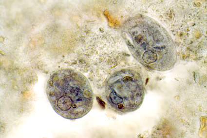
Iron-haematoxylin stain of trophozoites of Entamoeba coli
(note the "dirty" cytoplasm characteristic of E. coli).
Source: http://www.atlas-protozoa.com

At our lab we supplement microscopy of faecal concentrates with DNA-based detection of parasites. For some clinically significant parasites, we do a routine screen by PCR, since this is more sensitive than microscopy of faecal concentrates and because this is a semi-automated analysis that involves only DNA extraction, PCR and test result interpretation, which are all things that can be taught easily. Major drawbacks of diagnostic PCR is that you cannot really distinguish between viable (patent infection) and dead organisms (infection resolving, e.g. due to treatment). This is why, in the case of Blastocystis, you may want to do a stool culture as well (at least in post-treatment situations), since only viable cells will be able to grow, obviously.

Two diagnostic real-time PCR analyses have been published, one using CYBR Green and one using a TaqMan probe.

Now, it certainly differs from lab to lab as to which method is used for Blastocystis detection. Some labs apparently apply thresholds for number of parasites detected per visual field, and only score a sample positive if more than 5 parasites per visual field have been detected. I see no support for choosing a threshold, since 1) we do not know whether any Blastocystis-related symptoms are exacerbated by parasite intensity, 2) the number of parasites detected in a faecal concentrate may depend on so many things which have nothing to do with the observer (fluctuations in shedding for instance), and 3) the pathogenic potential of Blastocystis may very well depend on subtype.

If Blastocystis was formally acknolwedged as a pathogen, like Giardia, standardisation of methods would have happened by now. Meanwhile, we can only advocate for the use of PCR and culture if accurate diagnosis of Blastocystis is warranted, while permanent staining of fixed faecal samples constitutes a very good alternative in situations where PCR is not an option.

I have the impression that some labs do DNA-based detection of microbes, including protozoa, and that a result such as "taxonomy unknown" is not uncommon. I don't know how these labs have designed their molecular assays, and therefore I cannot comment on the diagnostic quality and relevance of those tests... it also depends on whether labs do any additional testing as well, such as the more traditional parasitological tests. However, we do know that there is a lot of organisms in our intestine, for which no data are available in GenBank, which is why it is sometimes impossible to assign a name to e.g. non-human eukaryotic DNA amplified from a stool sample.

* More than 1 billion people may harbour Blastocystis.
* Blastocystis is found mainly in the large intestine.
* 95% of humans colonised by Blastocystis have one of the following subtypes: ST1, ST2, ST3, ST4.
* DNA-based detection combined with culture ensures accurate detection of Blastocystis in stool samples and enables subtyping and viability assessment.


Further reading:

Poirier P, Wawrzyniak I, Albert A, El Alaoui H, Delbac F, & Livrelli V (2011). Development and evaluation of a real-time PCR assay for detection and quantification of blastocystis parasites in human stool samples: prospective study of patients with hematological malignancies. Journal of clinical microbiology, 49 (3), 975-83 PMID: 21177897

Röser D, Nejsum P, Carlsgart AJ, Nielsen HV, & Stensvold CR (2013). DNA of Dientamoeba fragilis detected within surface-sterilized eggs of Enterobius vermicularis. Experimental parasitology, 133 (1), 57-61 PMID: 23116599

Scanlan PD, & Marchesi JR (2008). Micro-eukaryotic diversity of the human distal gut microbiota: qualitative assessment using culture-dependent and -independent analysis of faeces. The ISME journal, 2 (12), 1183-93 PMID: 18670396

Stensvold CR, Ahmed UN, Andersen LO, & Nielsen HV (2012). Development and Evaluation of a Genus-Specific, Probe-Based, Internal-Process-Controlled Real-Time PCR Assay for Sensitive and Specific Detection of Blastocystis spp. Journal of clinical microbiology, 50 (6), 1847-51 PMID: 22422846

Stensvold CR, Arendrup MC, Jespersgaard C, Mølbak K, & Nielsen HV (2007). Detecting Blastocystis using parasitologic and DNA-based methods: a comparative study. Diagnostic microbiology and infectious disease, 59 (3), 303-7 PMID: 17913433

Stensvold CR, & Nielsen HV (2012). Comparison of microscopy and PCR for detection of intestinal parasites in Danish patients supports an incentive for molecular screening platforms. Journal of clinical microbiology, 50 (2), 540-1 PMID: 22090410