Showing posts with label culture. Show all posts

Friday, December 2, 2016

This Month in Blastocystis Research (NOV 2016)

Oftentimes, I receive emails from colleagues wanting to know how you subtype Blastocystis, how to grow them in culture, and how to freeze down cultures.

I'm very pleased to announce that Dr Graham Clark and I have developed protocols for exactly these activities and published them in Wiley's 'Current Protocols in Microbiology'; please go here for the subtyping protocol and here for the culture and cryopreservation protocols.
These should not only be seen as SOPs but also as a resource that enables standardization within the field.

Unfortunately, we have not yet come up with a protocol on how to axenise Blastocystis cultures, i.e., get rid of metabolically active organisms other than Blastocystis in cultures while keeping Blastocystis alive and multiplying.

We are well aware that many might not have access to these protocols because they haven't subscribed to Wiley Online Library; good news is that reprints will be available on request!


References:

Stensvold CR, & Clark CG (2016). Molecular Identification and Subtype Analysis of Blastocystis. Current Protocols in Microbiology, 43 PMID: 27858971  

Clark CG, & Stensvold CR (2016). Blastocystis: Isolation, Xenic Cultivation, and Cryopreservation. Current Protocols in Microbiology, 43 PMID: 27858970

Monday, April 29, 2013

'Invasive Blastocystis' in ECCMID 2013

ECCMID - the annual European Congress of Clinical Microbiology and Infectious Diseases (hosted by ESCMID) is currently taking place in Berlin. This year, I'm not attending, but I've been scanning the abstract book for 'Blastocystis', and it appears that an oral presentation was scheduled for yesterday in the "Emerging Infectious Diseases" section:

First of all: it's great to see fellow researchers screening larger (i.e. hundreds) of faecal DNAs by PCR for Blastocystis. I wish more people would do that to produce reliable data on prevalence and subtypes.

Now, as I've already mentioned, there are currently mainly two methods in use for subtyping, barcoding and STS PCR, and recently I evaluated these. To cut a long story short, barcoding is recommended for subtyping, since the STS method, which was used in the study by Tarasova et al. (abstract), appears to miss the majority of ST4 strains (the major genotype), and moreover, no STS primers exist for ST8 and ST9 (or any of the other 8 subtypes identified to date, but which have only been found in animals). So, the subtype data found in this study should be interpreted with this in mind.

Importantly however, I'm not sure whether the authors used the original Yoshikawa STS terminology or the terminology acknowledged in our 2007 consensus.

First, let us assume that consensus terminology is used. Then it's surprising to find ST5 in human samples in the first place, and finding a ST5 prevalence of 45% in a cohort of humans included in a larger study like this is very unlikely based on current evidence of more than 3,000 observations from all over the world, where the overall prevalence of ST5 in humans is <1%. Also, finding so much ST6 is also really striking. Also, if the consensus terminology is used, then I'm a bit puzzled why the authors put emphasis on ST7 not being found, since ST7 is relatively rare in humans.

And so let us assume that consensus terminology was not used, and the original Yoshikawa terminology was used instead. This would translate into STs 4, 6, and 7 not being detected in the CVH group. Which makes sense, since ST6 is extremely rare (at least in Europe), ST7 is only seen on occasion, and, as I said, the majority of ST4 infections are likely to go undetected by the STS method. However, ST4 appears quite common in Europe, and I suspect that it should be quite common in St Petersburg as well. But then there is one thing that comes to my mind: If ST4 infections are common, then there should be a relatively large number of samples detected by PCR which were untypable by PCR...and there is no information on untypable positive samples in the abstract...
But what is more:  STS subtype 5 translates into ST2 in consensus terminology, and similarly STS subtype 6 equals ST5 (yes, it may seem confusing, but we have provided a table in the 2007 consensus paper to make this easy). This means that no matter which of the two terminologies were used, ST5 is seen in abundance in patients with CVH in St Petersburg! Which is a very remarkable observation, and maybe more interesting than the rest of the data, which  I, by the way, find a bit difficult to follow (I expected to learn something about Blastocystis invasion, when I read the title of the abstract, but there is no data or information on invasiveness... and I'm very curious as to how the authors managed to obtain such a high number of samples from 'healthy people'! To evaluate the prevalence of Blastocystis in the control group, demographic data are needed, and a prevalence as low as 5.3% among healthy individuals makes me suspect that this control group consisted of newborns/toddlers who generally have a low prevalence of Blastocystis). Also, since when was ST1 'zoonotic'?

Anyway, often conference abstract are previews of upcoming articles, and so I expect that there will be a paper out soon from this group, and hopefully these issues will be clarified. The occasional confusion in Blastocystis epidemiology could be reduced to a minimum if everyone got into using barcoding and the Blastocystis 18S subtyping site (and go here for a video introduction to Blastocystis subtyping).

Are some citizens of St Petersburg infected by Blastocystis sp. ST5, a subtype seen primarily in livestock and African apes? Source

References:
Tarasova E, Suvorova M, Sigidaev A, Suvorov A. Blastocystis invasion in patients with chronic viral hepatitis in Saint Petersburg. ECCMID 2013 abstract O338.

Alfellani MA, Stensvold CR, Vidal-Lapiedra A, Onuoha ES, Fagbenro-Beyioku AF, & Clark CG (2013). Variable geographic distribution of Blastocystis subtypes and its potential implications. Acta Tropica, 126 (1), 11-8 PMID: 23290980

Stensvold CR (2013). Comparison of sequencing (barcode region) and sequence-tagged-site PCR for Blastocystis subtyping. Journal of Clinical Microbiology, 51 (1), 190-4 PMID: 23115257

Stensvold CR, Suresh GK, Tan KS, Thompson RC, Traub RJ, Viscogliosi E, Yoshikawa H, & Clark CG (2007). Terminology for Blastocystis subtypes--a consensus. Trends in Parasitology, 23 (3), 93-6 PMID: 17241816

Friday, February 22, 2013

Bubbly Blasto!

Yesterday, I was checking up on a fresh Blastocystis culture. I loaded 20 µL of the culture "sediment" on to a glass slide, placed the cover slip on top and examined it by light microscopy. While examining the slide, I observed a multitude of dividing cells, indicating vigorous growth and a thriving strain, and once again I was struck by the appearance of dividing Blastocystis. This is basically what they may look like:

Like soap bubbles really, only a lot smaller obviously (mikrons), and somewhat opaque! You'll see them in different sizes and the way they divide looks just like this. Apparently some sort of random budding or multiple fission. You'll see little more than this bubbly structure, which means that there are very few morphological hallmarks to describe. A few nuclei may be discernible along the cytoplasmatic rim, but that's about it when you use light microscopy. Ultrastructural and biochemical analysis is required if you hope to be able to describe some of the processes involved in reproduction.

We often say that Blastocystis organisms representing different subtypes are morphologically indistinguishable; what this actually means is that we do not have the tools to differentiate them morphologically. There may actually be great variation between strains in terms of for instance how they grow in vivo and in vitro and maybe also how they reproduce. Vacuolar forms are the most common form seen in xenic cultures, but other morphotypes are sometimes observed, for instance the granular stage, which, in my experience, is typically seen in cultures that are not “well looked after”, i.e. where medium is not being replaced about twice a week. Dunn and colleagues. (1989) observed that the granular stage could arise from vacuolar stages in cultures where the concentration of horse serum was increased.

I have previously stated that there is no evidence for phagocytosis in Blastocystis. Actually, Dunn et al. (1989) captured what they thought to be bacterial engulfment by ultra-structural analysis, and they also observed bacteria-engulfing pseudopodia in amoeboid stages, in which degraded bacteria were observed. I don't think that I've ever come across this amoeboid stage, but it has been described by quite a few researchers.

Anyway, let's hope for another kind of bubbles this Friday night!

Suggested reading:

Dunn LA, Boreham PF, & Stenzel DJ (1989). Ultrastructural variation of Blastocystis hominis stocks in culture. International Journal for Parasitology, 19 (1), 43-56 PMID: 2707962

Monday, January 14, 2013

A Penny For Your Thoughts

So, what should we do about Blastocystis? What do we want to know?

I believe the imminent answer to the latter question is easy: We want to know whether it’s pathogenic, whether we should treat it and how. But I also think that there are many other interesting aspects of Blastocystis which are also of broad interest to the general public, namely: How about the many cases of asymptomatic Blastocystis carriage? What does Blastocystis do in our guts? Could it have any potentially beneficial impact on our health?

Given the fact that Blastocystis has not been implicated in any outbreaks (admittedly: I guess that no one actually ever looked for Blastocystis in outbreak investigations... except for me!), I reckon that the chance of it being involved in acute diarrhoea is small. So, in that respect it's very different from the other intestinal protists such as Giardia, Cryptosporidium, Cyclospora, microsporidia, even Entamoeba histolytica. It's actually more reminiscent of helminth infections, which are are often chronic, and when light hardly give rise to symptoms (depending on species that is!).So I'm more thinking along the lines of co-evolution, adaptation, etc.

Maybe future research will call for a shift in paradigm, but until then I think that we should do what we already can, just at a larger scale and see where it takes us, namely:

Saturday, November 10, 2012

How Hard Can It Be?


How strange the world of clinical microbiology is when you compare the fields of mycology, parasitology, bacteriology and virology to each other. Such different possibilities, opportunities, limitations, and diagnostic challenges! The 3 month mortality rate of invasive aspergillosis, a disease mainly caused by Aspergillus fumigatus and seen in mainly patients with haematological malignancies, patients undergoing allogenic HSCT and patients in ICUs, may be as high as 60%, and therefore a quick and reliable diagnosis is mandatory to secure timely therapeutic intervention. But, - Aspergillus fumigatus happens to be ubiquitous, and contamination of patient samples, whether blood or airway samples, may always be a potential cause of false-positive test results, and one of the reasons why the use of PCR as a first line diagnostic tool in routine mycology labs is still limited. Antigen tests, such as the Galactomannan antigen test, which also allow quick diagnosis can also be false-positive, not only due to sample contamination, but also due to galactomannan residues in medical compounds, such as the widely applied antibiotic Tazocin (piperacillin-tazobactam), which means that patients who have been given this drug and who submit a blood sample for galactomannan testing may test slightly positive even in the absence of an Aspergillus infection.
These are only some classical examples. In the field of mycology, positive predictive values (PPV; i.e. what is the probability of disease given a positive test result) are sometimes unacceptably low, and the lower the prevalence of the disease, the lower the PPV. This means that you need a lot of experience and knowledge on pre-test-probability + data from clinical and diagnostic work-ups, including anamnestic details, to determine whether or not the patient should receive therapy, such as treatment with voriconazole, -  a relatively expensive drug.

Aspergillus fumigatus - the most common cause of invasive aspergillosis - on blood agar.

In the parasitology lab, however, things are quite different. Contamination of patient samples is rarely an issue, and in most cases not possible at all (disregarding DNA contamination of course). Specificity of microscopy is very often very high (close to 100%), which means that the PPV is very high even in cases where the disease is rare. Hence, if cysts of Giardia have been detected in your stool, it's due to the presence of the parasite in your body. It's a bit more tricky with PCR-based analyses, where the specificity does not rely on your ability to visually distinguish between e.g. Giardia and non-Giardia elements, but where it's all about designing oligos that anneal only to Giardia-DNA.
While in the mycology lab we struggle with low PPVs, one of the biggest challenges for me and my colleagues in the parasitology lab is to optimise the negative predictive value (NPV) of a faecal parasite diagnostic work-up - how can we rule out parasitic disease by cost-effectively putting together a panel of as few tests as possible?

There are many other differences. For instance, you can grow bacteria and fungi in the lab very easily, in fact, culture of bacteria and fungi is an essential diagnostic tool, which also allows you to submit the strain to antibiotic or antimycotic susceptibility testing and molecular characterisation/MALDI-TOF analysis in case you are not sure about the species ID. So, you have the strains right there in front of you, on agar plates, and they grow and grow, and you can keep them for as long as you like, - clean, non-contaminated strains on selective media.
You can't really do that with parasites, not nearly to the same extent and as easily, that is. For instance, you can culture Blastocystis directly from stool for sure (go here for the protocol), but only in the presence of bacteria (some of my colleagues do actually now and then manage to grow strains of Blastocystis in the absence of bacteria, they obtain what is called "axenic" cultures, but I believe that they cannot do it consistently and in limited time.). And it's a pity, since there is so much you can do when you have "clean" patient strains. Apart from susceptibility testing (which would actually be a bit difficult since Blastocystis is strictly anaerobic, so you can't really have it in microtiter plates or on RPMI plates on the table in front of you, but the strains could be challenged in the growth tubes), you can also extract DNA, and you would know that all the DNA that you extract from the isolate is from that particular strain, and not from bacterial contaminants. You can use the strain for production of antigens which can be used in ELISAs and used to generate mono- and polyclonal antibodies... Sequencing genomes of various subtypes would be a lot easier and quicker, and so on...

So, what appears obvious in one field of microbiology is not as obvious in another field, and vice versa. I wish Blastocystis was much easier to isolate. Dientamoeba too. Dientamoeba is probably as common as Blastocystis, and not rarely seen in co-infections. It is strange to contemplate that a parasite infecting hundreds of millions of people has not yet had its genome sequenced? We have no clue when it comes to effector proteins in Dientamoeba, and also for this parasite, what we know about its clinical significance relies mainly on epidemiological data.

There is no doubt that concerted efforts of experienced scientists should make it possible to develop appropriate and relevant culture protocols for these parasites. It does, however, require a lot of resources and time to get to know these common, but oh so fragile and reclusive little creatures...

Further reading:
Clark CG, & Diamond LS (2002). Methods for cultivation of luminal parasitic protists of clinical importance. Clinical microbiology reviews, 15 (3), 329-41 PMID: 12097242

Verweij PE, Kema GH, Zwaan B, & Melchers WJ (2012). Triazole fungicides and the selection of resistance to medical triazoles in the opportunistic mould Aspergillus fumigatus. Pest management science PMID: 23109245

Stensvold, C., Jørgensen, L., & Arendrup, M. (2012). Azole-Resistant Invasive Aspergillosis: Relationship to Agriculture Current Fungal Infection Reports, 6 (3), 178-191 DOI: 10.1007/s12281-012-0097-7

Maertens J, Theunissen K, Verhoef G, & Van Eldere J (2004). False-positive Aspergillus galactomannan antigen test results. Clinical infectious diseases : an official publication of the Infectious Diseases Society of America, 39 (2), 289-90 PMID: 15307045
 
Munasinghe VS, Stark D, & Ellis JT (2012). New advances in the in-vitro culture of Dientamoeba fragilis. Parasitology, 139 (7), 864-9 PMID: 22336222

Tuesday, July 10, 2012

Blastocystis Culture in Jones' Medium

Upon request I have now posted the protocol on one of the simplest media used for Blastocystis culture, Jones' Medium, - please go to the tab (page) "Lab Stuff".

You can read about Blastocystis culture in some of my other blog posts, use the search box or the labels feature.

Please be aware that this is for xenic culture only - i.e. culture in the presence of bacteria. It's quick, inexpensive, very reliable (at least for human samples) and isolates can be kept this way for months/years - all you need is an incubator.

Extracting DNA from cultures and using it for subtyping usually yields excellent results.

I have never tried to cryopreserve (freeze down) Blastocystis using Jones' Medium, but it is possible (at least when Robinson's Medium is used).

More reading:

Stensvold CR, Arendrup MC, Jespersgaard C, Mølbak K, & Nielsen HV (2007). Detecting Blastocystis using parasitologic and DNA-based methods: a comparative study. Diagnostic microbiology and infectious disease, 59 (3), 303-7 PMID: 17913433

And, if you are interested in culture of intestinal protists in general, why not look up

Clark CG, & Diamond LS (2002). Methods for cultivation of luminal parasitic protists of clinical importance. Clinical microbiology reviews, 15 (3), 329-41 PMID: 12097242

Saturday, July 7, 2012

Blastocystis Nutrition

A reader of this blog asked me about the nutritional requirements of Blastocystis and whether I thought the parasite can be eradicated by fasting.

Given my background (I'm not a dietitian for starters), I guess my best way of approaching this is by drawing on my experience from the lab. When we diagnose Blastocystis, we have multiple methods to choose from, some of which are better than others (please look up previous posts here for more information). Short term (i.e. 24-48 h) in-vitro culture at 37 °C in Jones' medium is almost as sensitive as PCR (molecular detection). This means that if viable Blastocystis is present in a faecal sample, then it will most probably "come up" in culture, which means that in a day or two, we will be able to detect those "characteristically non-characteristic" soap bubble structures (the vacuolar stage) by light microscopy of a small portion of the culture - they will be all over the place!

So, what's Jones' medium? Well, Blastocystis can be cultured in a variety of different media, some of which are very primitive. Jones' medium is probably one of the simplest media, and consists mainly of electrolytes, yeast extract (contains nucleic acids) and horse serum (containing lipids). Importantly, we don't even have to add starch to the medium, when we culture Blastocystis xenically (i.e. under non-sterile conditions and this is what we always do when using culture diagnostically). Blastocystis has also been grown in a saline-serum medium, again in the presence of bacteria.

Apart from providing the anaerobic environment required for Blastocystis to thrive, bacteria most probably constitute a significant source of nutrients for the parasite. We can consistently keep strains of Blastocystis in xenic culture for weeks, months, years, observing vigorous growth, and it is clear that the bacteria and the simple medium supply nutrients in abundance. I have never managed to axenise (i.e. eliminate bacteria from) a culture, but others have been successful at times. One of the pioneers in Blastocystis research, Charles H. Zierdt, noted that the axenisation of Blastocystis usually takes weeks/months with a continuous reduction of bacterial numbers and species, until one species, usually a Bacteroides sp., remains; elmination of the last bacterial species may or may not result in axenisation, simply depending on the need for bacterial support. One of our future goals is to characterise the bacterial flora in individuals with and without Blastocystis.

I believe that even during fasting, Blastocystis will have plenty of access to essential nutritional components. It is possible that fasting may impact the intestinal bacterial flora, and if Blastocystis is dependent on a certain bacterial flora, it may be so that the parasite can be "manipulated" by manipulating the intestinal flora.

Useful reading:

Clark CG, & Diamond LS (2002). Methods for cultivation of luminal parasitic protists of clinical importance. Clinical microbiology reviews, 15 (3), 329-41 PMID: 12097242
 
Zierdt CH (1991). Blastocystis hominis--past and future. Clinical microbiology reviews, 4 (1), 61-79 PMID: 2004348

Sunday, July 1, 2012

Do I Get Diagnosed Correctly?

I can tell especially from Facebook discussions that people across the globe wanting to know about their "Blastocystis status" are worried that they are receiving false-negative results from their stool tests, and that many Blastocystis infections go unnoticed. And I think I should maybe try and say a few things on this (please also see a recent blog post on diagnosis, - you'll find it here). I might try and simplify things a bit in order not to make the post too long.

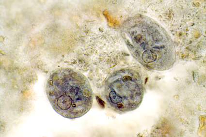
Below, you'll find a tentative representation of the life cycle of Blastocystis. It is taken from CDC, from the otherwise quite useful website DPDx - Laboratory Identification of Parasites of Public Health Concern.

Proposed life cycle of Blastocystis.
 
I don't know how useful it is, but what's important here is the fact that we accidentally ingest cysts of Blastocystis, and we shed cysts that can be passed on to other hosts. The cyst stage is the transmissible stage, and the way the parasite can survive outside the body; we don't know for how long cysts can survive and remain infective. In our intestine and triggered by various stimuli, the cysts excyst, transiting to the non-cyst form, which could be called the trophozoite / "troph" stage, or to use a Blastocystis-specific term, the "vacuolar stage" (many stages have been described for Blastocystis, but I might want to save that for later!). This is possibly the stage in the life cycle where the parasite settles, thrives, multiplies, etc. You can see a picture of vacuolar stages in this blog post. Many protozoa follow this simple life cycle pattern, among them Giardia and most species of Entamoeba. If the stool is diarrhoeic and you are infected by any one or more of these parasites, it may be so that only trophozoites, and, importantly, no cysts, are shed! This has something to do with reduced intestinal transit time and, maybe more importantly, the failure of the colon to resorb water from the stool which means that the trophozoites do not get the usual encystation stimuli. Importantly, trophozoites are in general non-infectious.

There is documentation that once colonised with Blastocystis, you may well carry it with you for years on end, and as already mentioned a couple of times, no single drug or no particular diet appears to be capable of eradicating Blastocystis - this is supported by the notion that Blastocystis prevalence seems to be increasing by age, although spontaneous resolution may not be uncommon, - we don't know much about this. Now, although day-to-day variation in the shedding of Blastocystis has been described, it is my general impression that colonised individuals may shed the parasite with each stool passage, and well-trained lab technicians/parasitologists will be able to pick up Blastocystis in a direct smear (both cysts and trophs may be seen). To do a direct smear you simply just mix a very small portion of the stool with saline or PBS on a slide, put a cover slip over it and do conventional light microscopy at x200 (screening) or x400 (verification). Very light infections may be difficult to detect this way, and if you don't have all the time in the world, a direct smear may not be the first choice.

The "king" of parasitological methods, however, is microscopy of faecal concentrates (Formol Ethyl Acetate Concentration Technique and any variant thereof), which is remarkable in its ability to detect a huge variety of parasites. Especially cysts of protozoa (e.g. Giardia and Entamoeba) and eggs of helminths (e.g. tapeworm, whipworm and roundworm) concentrate well and are identified to genus and species levels based on morphology. The method is not as sensitive as DNA-based methods such as PCR, but as I said, has the advantage of picking up a multitude of parasites and therefore good for screening; PCR methods are targeted towards particular species (types) of parasites. A drawback of the concentration method is that it doesn't allow you to detect trophzoites (i.e. the fragile, non-cystic stage), and, as mentioned, diarrhoeic samples may contain only trophozoites and no cysts...

In many countries it is very common for people to be infected by both protozoa and helminths, and in those countries microscopy of faecal concentrates is a relevant diagnostic choice. In Denmark and many Western European countries, the level of parasitism is higher than might be expected (from a hygiene and food safety point of view) but due to only few parasitic species. Paradoxically, the intestinal parasites that people harbour in this part of the world are parasites that do not concentrate well. They are mainly:

1) Blastocystis
2) Dientamoeba fragilis
3) Pinworm (Enterobius vermicularis)

Only troph stages have been described for Dientamoeba fragilis and it may be transmitted by a vector, such as pinworm (look up paper by Röser et al. in the list below for more information); this mode of transmission is not unprecedented (e.g. Histomonas transmission by Heterakis). Eggs of pinworm may be present in faeces, but a more sensitive method is the tape test.

Now, Blastocystis often disintegrates in the faecal concentration process, and while you might be lucky to pick up the parasite in a faecal concentrate, you shouldn't count on it, and hence the method is not reliable, unless the faecal sample was fixed immediatley after being voided. This is key, and also why fixatives are used for the collection of stool samples in many parts of the world - to enable the detection of fragile stages of parasites. There are many fixatives, e.g. SAF (sodium acetate-acetic formalin), PVA (poly-vinyl alcohol) and even plain formalin will do the trick if it's just a matter of preserving the parasite in the sample. If SAF or PVA is used, this allows you to do permanently stained smears of faecal concentrates, and you will be able to pick up not only cysts of protozoa, but even trophozoites. Trichrome and iron-haematoxylin staining are common methods and are sensitive but very time-consuming and may be related to some health hazards as well due to the use of toxic agents. But this way of detecting parasites is like good craftmanship - it requires a lot of expertise, but then you get to look at fascinating structures with intriguing nuclear and cytoplasmatic diagnostic hallmarks. Truly, morphological diagnosis of parasites is an art form! Notably, samples preserved in such fixatives may be useless for molecular analyses.

Iron-haematoxylin stain of trophozoites of Entamoeba coli
(note the "dirty" cytoplasm characteristic of E. coli).
Source: http://www.atlas-protozoa.com

At our lab we supplement microscopy of faecal concentrates with DNA-based detection of parasites. For some clinically significant parasites, we do a routine screen by PCR, since this is more sensitive than microscopy of faecal concentrates and because this is a semi-automated analysis that involves only DNA extraction, PCR and test result interpretation, which are all things that can be taught easily. Major drawbacks of diagnostic PCR is that you cannot really distinguish between viable (patent infection) and dead organisms (infection resolving, e.g. due to treatment). This is why, in the case of Blastocystis, you may want to do a stool culture as well (at least in post-treatment situations), since only viable cells will be able to grow, obviously.

Two diagnostic real-time PCR analyses have been published, one using CYBR Green and one using a TaqMan probe.

Now, it certainly differs from lab to lab as to which method is used for Blastocystis detection. Some labs apparently apply thresholds for number of parasites detected per visual field, and only score a sample positive if more than 5 parasites per visual field have been detected. I see no support for choosing a threshold, since 1) we do not know whether any Blastocystis-related symptoms are exacerbated by parasite intensity, 2) the number of parasites detected in a faecal concentrate may depend on so many things which have nothing to do with the observer (fluctuations in shedding for instance), and 3) the pathogenic potential of Blastocystis may very well depend on subtype.

If Blastocystis was formally acknolwedged as a pathogen, like Giardia, standardisation of methods would have happened by now. Meanwhile, we can only advocate for the use of PCR and culture if accurate diagnosis of Blastocystis is warranted, while permanent staining of fixed faecal samples constitutes a very good alternative in situations where PCR is not an option.

I have the impression that some labs do DNA-based detection of microbes, including protozoa, and that a result such as "taxonomy unknown" is not uncommon. I don't know how these labs have designed their molecular assays, and therefore I cannot comment on the diagnostic quality and relevance of those tests... it also depends on whether labs do any additional testing as well, such as the more traditional parasitological tests. However, we do know that there is a lot of organisms in our intestine, for which no data are available in GenBank, which is why it is sometimes impossible to assign a name to e.g. non-human eukaryotic DNA amplified from a stool sample.

* More than 1 billion people may harbour Blastocystis.
* Blastocystis is found mainly in the large intestine.
* 95% of humans colonised by Blastocystis have one of the following subtypes: ST1, ST2, ST3, ST4.
* DNA-based detection combined with culture ensures accurate detection of Blastocystis in stool samples and enables subtyping and viability assessment.


Further reading:

Poirier P, Wawrzyniak I, Albert A, El Alaoui H, Delbac F, & Livrelli V (2011). Development and evaluation of a real-time PCR assay for detection and quantification of blastocystis parasites in human stool samples: prospective study of patients with hematological malignancies. Journal of clinical microbiology, 49 (3), 975-83 PMID: 21177897

Röser D, Nejsum P, Carlsgart AJ, Nielsen HV, & Stensvold CR (2013). DNA of Dientamoeba fragilis detected within surface-sterilized eggs of Enterobius vermicularis. Experimental parasitology, 133 (1), 57-61 PMID: 23116599

Scanlan PD, & Marchesi JR (2008). Micro-eukaryotic diversity of the human distal gut microbiota: qualitative assessment using culture-dependent and -independent analysis of faeces. The ISME journal, 2 (12), 1183-93 PMID: 18670396

Stensvold CR, Ahmed UN, Andersen LO, & Nielsen HV (2012). Development and Evaluation of a Genus-Specific, Probe-Based, Internal-Process-Controlled Real-Time PCR Assay for Sensitive and Specific Detection of Blastocystis spp. Journal of clinical microbiology, 50 (6), 1847-51 PMID: 22422846

Stensvold CR, Arendrup MC, Jespersgaard C, Mølbak K, & Nielsen HV (2007). Detecting Blastocystis using parasitologic and DNA-based methods: a comparative study. Diagnostic microbiology and infectious disease, 59 (3), 303-7 PMID: 17913433

Stensvold CR, & Nielsen HV (2012). Comparison of microscopy and PCR for detection of intestinal parasites in Danish patients supports an incentive for molecular screening platforms. Journal of clinical microbiology, 50 (2), 540-1 PMID: 22090410

Wednesday, April 18, 2012

Blastocystis Subtyping in Routine Microbiology Labs

When I speak to colleagues in and outside Europe and visit research portals and social media, including Facebook groups, I get the impression that Blastocystis subtyping is something that is still very rarely done, despite the fact that most clinical microbiologists and biologists acknowledge that subtypes may differ in terms of clinical significance and in other respects. We get new data on Blastocystis subtypes in various cohorts from time to time from research groups around the world, but all reports are characterised by relatively small sample sizes and subtyping methodology has not yet been standardised. This type of research is moreover challenged by the fact that Blastocystis is common in healthy individuals (i.e. people not seeing their GPs for gastrointestinal problems), and this makes it extremely difficult to identify the subtype distribution in the "background" population.

Although we recommend barcoding (see one of my previous posts) as the subtyping method of choice, there is no "official report" identifying the Blastocystis subtyping gold standard. Therefore, I'm currently setting up a lab project that is going to help us compare the most common methods used for subtyping in order to identify the one most suitable. I emphasise that the best method used for subtyping is not the PCR that should be used for diagnostic purposes, mostly due to the fact that PCRs for subtyping amplify 300-600 bp, which are much longer amplicons than the one we go for in diagnostic PCRs (typically 80-100 bp). We therefore recommend our novel TaqMan-based real-time PCR for initial diagnosis, or culture, which is inexpensive and relatively easy and provides you with a good source of cells for DNA extraction.
I hope that we will be able to come up with some robust data soon that will allow us to recommend the most suitable approach and hope to publish our results in a clinical microbiology journal of high impact, and I hope that this will prompt Blastocystis subtyping in many labs. Once this report has been published, I intend to upload a protocol here at the site where lab procedures for diagnosis and subtyping will be described in detail. Stay tuned!

Sunday, April 8, 2012

A Few Words On Blastocystis Morphology and Diagnosis

Blastocystis is a sinlge-celled parasite. The parasite produces cysts (probably the transmissible form) and vegetative stages (including the stage commonly referred to as the vacuolar stage). Vegetative stages are commonly seen in fresh faecal samples and in culture. This is what they look like under light microscopy:

Vegetative stages of Blastocystis (unstained) (source: www.dpd.cdc.gov)


Using permanent staining of fixed faecal material, the eccentrically located nuclei become more apparent:

Vegegtative stages of Blastocystis (Trichrome stain) (source: www.dpd.cdc.gov)


Although sensitive, permanent staining techniques (e.g. Trichrome, Giemsa and Iron Haematoxylin) are relatively time-consuming, impractical and expensive. Since also conventional concentration of unfixed stool using e.g. the Formol Ethyl-Acetate Concentration Technique is not appropriate for diagnosis (Blastocystis cysts are very difficult to pick up, and vacuolar stages become distorted or disintegrate), we recommend short-term in-vitro culture (using Jones' or Robinson's medium) and/or Real-Time-PCR on genomic DNAs extracted directly from faeces using QIAGEN Stool Mini Kit (QIAGEN, Hilden, Germany) or - in modern laboratories - by automated DNA extraction robots. Once genomic DNAs have been extracted and screened by PCR, positive samples can be submitted to subtyping using the barcoding method, and DNAs can be screened for other parasites by PCR as well. In fact the use of insensitive methods to distinguish carriers from non-carriers is one of our greatest obstacles to obtaining valid prevalence data on Blastocystis.

Having an isolate in culture adds the benefit of having a continuous source of DNA for further genetic characterisation (for instance complete SSU-rDNA sequencing) in case a particular isolate turns out to be genetically different from those already present in GenBank or the isolate database at Blastocystis Sequence Typing Home Page. And chances are that there are quite a few "novel" subtypes out there... especially in animals. However, Blastocystis from animals may not always be successfully established in culture.