Friday, June 21, 2013

This Month In Blastocystis Research (JUN 2013)

Another paper in the string of publications coming out from the PhD study by Dr Alfellani (London School of Hygiene and Tropical Medicine) has just appeared in PubMed.

Dr Alfellani and his colleagues have done a great job in analysing a multitude of samples from humans, non-human primates and animals; I have previously blogged about their observations from studies of human and non-human primates. Moreover, they have surveyed available data in order to better discuss their own findings, and the work has contributed significantly to what today is known about the host specificity, genetic diversity, phylogeography and general molecular epidemiology of Blastocystis.

Alfellani's most recent paper is published in the journal Protist, and it deals with the 'Genetic Diversity of Blastocystis in Livestock and Zoo Animals'.

It is quite a large paper which includes a lot of new information and a comprehensive (and hopefully exhaustive) table summarising Blastocystis subtype data in all relevant hosts (humans, non-human primates, other mammals and birds).

I will highlight a couple of things from the paper:

1. Apart from reporting on virtually complete SSU rDNA sequences from a couple of subtypes for which entire SSU rDNA sequences have yet not been available, we also report on three novel subtypes. Until recently, we only knew about 14 subtypes (ST1-ST14), of which ST1-ST9 can be found in humans. Now, three additional subtypes have been identified; ST15 in artiodactyls (camel and sheep) and non-human primates (chimpanzee and gibbon), ST16 in kangaroos, and ST17 in gundis.

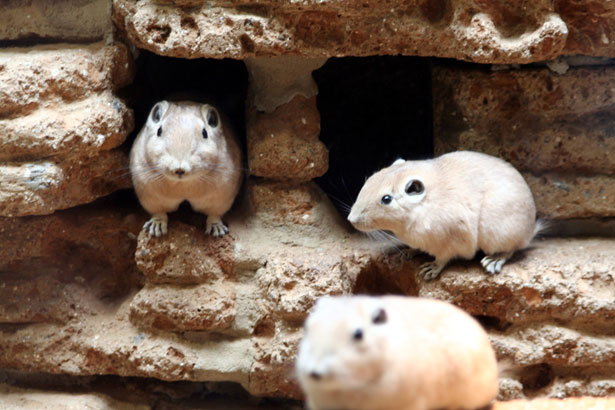
The Gundi (Ctenodactylus gundi) is a rodent living mainly in the deserts of Northern Africa. (Source)

2. Novel data arising from analysis of faecal samples from humans and animals in Sebha, Libya, strongly indicate that humans and animals in this area are infected by different subtypes: Humans appear to carry ST1, ST2, and ST3, while synanthropic animals (artiodactyls in this case) mostly have ST5 and ST10 infections, suggesting that livestock is not a major contributor to human Blastocystis infection.

To this end, there is growing evidence of quite a substantial degree of host specificity of Blastocystis.  Even when subtypes overlap between humans and animals, we have accumulating evidence that the strains found in humans and animals are different. This means that the hypothesis that animals constitute an important reservoir of human Blastocystis infections currently has very limited support. It is my clear impression that when a strain of ST6 or ST8 is detected in humans, this strain has most probably been transmitted from an animal source. But we very rarely see these subtypes in humans, at least in Europeans.

It will be extremely interesting to see how the universe of Blastocystis subtypes unfolds... by genetically characterising strains in humans and non-human hosts, we are building up a clearer picture of transmission patterns and evolutionary biology, including our adaptation to Blastocystis, and the parasite's adaptation to us and other hosts.

It is noteworthy that we are starting to see different subtypes in rodents. We have previously thought that generally, rodents were infected by ST4. But now we know that many rodents are not infected, and we also know that rodents may harbour subtypes other than ST4.

So,17 subtypes of Blastocystis are now known. We have probably only seen the top of the iceberg, since many host species have not yet been sampled from, and it is likely that we will see quite a few STs being identified in the nearest future. To this end it is necessary to have a consensus regarding the identification of novel subtypes. Along with the Protist paper we have uploaded a supplementary file (Appendix A, TXT format) with aligned reference sequences that can be used for phylogenetic analysis,  hoping that it will be useful to our colleagues. In a future blog post I will try to address the issues of identifying new subtypes more specifically.

ST15 is one of the more interesting subtypes since it appears to have quite a low host specificity - infecting both non-human primates and artiodactyls. Yet, we have come across it only now. ST15 and ST17 are remarkable in the way that they appear to be closer related to herptile and arthropod lineages, respectively, than to lineages from mammals.

Please note that virtually complete sequences of ST10, ST13, ST14, ST15, and ST17 analysed in the study have been released in GenBank just now.

Further reading:

Alfellani MA, Taner-Mulla D, Jacob AS, Imeede CA, Yoshikawa H, Stensvold CR, & Clark CG (2013). Genetic Diversity of Blastocystis in Livestock and Zoo Animals. Protist, 164 (4), 497-509 PMID: 23770574

Alfellani MA, Stensvold CR, Vidal-Lapiedra A, Onuoha ES, Fagbenro-Beyioku AF, & Clark CG (2013). Variable geographic distribution of Blastocystis subtypes and its potential implications. Acta Tropica, 126 (1), 11-8 PMID: 23290980

Alfellani MA, Jacob AS, Perea NO, Krecek RC, Taner-Mulla D, Verweij JJ, Levecke B, Tannich E, Clark CG, & Stensvold CR (2013). Diversity and distribution of Blastocystis sp. subtypes in non-human primates. Parasitology, 140 (8), 966-71 PMID: 23561720

Saturday, June 15, 2013

Blastocystis - 'Monsters Inside Me'

I woke up this morning, grabbed my smartphone and went on to check my Blastocystis Google Alert. There was one entry, and this was the one:


Now, I could probably do a post with hundreds of examples showing how the internet abounds with material that may misguide/misinform people on Blastocystis pathogenicity. As such, this video is a nice example of how you can diligently manipulate people into thinking that severe, debilitating disease can be caused by Blastocystis.

Now, before I move on, I have to say that if this is a documentary, I'm very sorry for the couple in this video who have suffered the pain and consequences of sudden debilitating illness. Precautions have to be taken when you are exposed to sewage to avoid contracting infections.

The symptoms that are described in the video - including the weight loss - could be attributable to many different bacterial and viral pathogens, even parasites such as Cryptosporidium and maybe also Giardia; to this end, the video provides us with no information on other pathogens found in the patient's stool. Even in the event that Blastocystis was the only potential pathogen found, other pathogens may have been overlooked if sensitive diagnostics were not taken into use.

It is possible that Bill Wilson contracted Blastocystis only after signing on to his plumbing contract, but it is also possible that he had it a long time before. Many of us (up to 30% of the healthy Danish population) are colonised, and colonisation is often chronic.

We are informed that the patient receives a course of metronidazole, a drug that is often used to treat Blastocystis, but which in fact has a limited efficacy in vivo when used alone. Bill apparently clears his symptoms after metronidazole treatment, but we do not know whether in fact Bill also clears his Blastocystis infection, which could be determined by post-treatment stool tests. Metronidazole is capable of clearing a large number of anaerobic bacterial and protozoan species, and it is not unlikely that the drug has eradicated one or more pathogens that Bill could have contracted during his work (or elsewhere), and so symptom relief may be due to clearance of a non-Blastocystis pathogen instead.

Finally, it may be so that symptom disappearance coincides with spontaneous pathogen resolution. Cryptosporidiosis, for instance, can cause quite debilitating disease even in immunocompetent individuals, causing the infected individuals to lose a lot of water due to diarrhoea lasting for days or even weeks, but the disease is usually self-limiting.

So, this video tells a story that makes the audience automatically think that Bill Wilson's disease is due to Blastocystis. Apart from the statement 'Complications from a Blastocystis hominis infection can be fatal' and the explanation of how metronidazole works on Blastocystis, there is not really any statements or information in the video that do not make sense; the video is just put together in a way so that the viewer automatically deduces that Blastocystis is the culprit. A diligent act of manipulation!

Please note that this post is about how information on Blastocystis can be conveyed to an audience and not about the particular case as such.

Reference:

Stensvold CR, Smith HV, Nagel R, Olsen KE, & Traub RJ (2010). Eradication of Blastocystis carriage with antimicrobials: reality or delusion? Journal of clinical gastroenterology, 44 (2), 85-90 PMID: 19834337

Saturday, May 25, 2013

This Month in Blastocystis Research (MAY 2013)

Now, we have a situation. Last month, I came up with the idea of the post series 'This Month in Blastocystis Research' developed for discussing a couple of papers on Blastocystis appearing recently in pubmed. However, this month only one Blastocystis release has emerged. It is in Turkish with an English abstract and so I'm not in the optimum position to review it. Overall, I'm not entirely clear on why the authors have chosen to publish the work. The paper is apparently about PCR amplification of Blastocystis specific DNA (using the barcoding primers) with subsequent cloning with a view to producing subtype information that could have been obtained simply by direct sequencing. At least when the goal is to subtype a particular positive sample, PCR + sequencing should suffice. Obviously, if you want to explore intra-subtype diversity, cloning is very useful. But it is time consuming for subtyping and also expensive. Therefore, for plain subtyping, I recommend the protocol that I put out on youtube a few weeks ago. The phylogenetic tree produced by the authors looks unfamiliar to me in that the clustering of the subtypes is quite different from the phylogenies inferred by other groups; this should not have anything to do with the SSU rDNA region explored; rather it may boil down to issues with alignment editing or the algorithm chosen for phylogenetic analysis. Well, we should be looking forward to more subtype data from Turkey! Incidentally, I was once involved in a Turkish study where we found ST1, ST2 and ST3 mainly, while ST4 was rare.

Since there are no other papers to discuss, I will try and compensate by providing a link to 'This Week in Parasitism' hosted by Vincent Racaniello and Dickson Despommier, who are going through a case of Blastocystis possibly contracted abroad during a field trip to Bali. Now, there's a lot of digression in this pod cast (some of which is actually quite enjoyable). Also, I do not agree with all the things said about Blastocystis in this conversation. If you cannot make the link work, you can access the podcast directly here

I do think it's a bit strange though that given the clinical focus of the talk, there is not a single word on paromomycin. But I guess the overall take home message is that treating Blastocystis is really difficult, and no single type of therapeutic intervention is consistently efficacious. Unfortunately, the two gentlemen do not touch upon the genetic diversity of Blastocystis, which is probably one of the most interesting things about Blastocystis currently known, and which may also be part of the reason why no single treatment modality seems to work every single time.


I wonder whether Blastocystis will always be stuck in shades of grey... or whether at some point we'll be able to make some clear-cut conclusions that will be useful for clinicians and clinical microbiologists...? I hope! And I believe we are certainly on our way!

Anyway, enjoy a bit of Blastocystis causerie!

Suggested reading:
Sakalar C, Uyar Y, Yürürdurmaz MA, Tokar S, Yeşilkaya H, Gürbüz E, Kuk S, & Yazar S (2013). [Cloning of Blastocystis sp Subtype 3 Small-subunit Ribosomal DNA]. Turkiye Parazitolojii Dergisi / Turkiye Parazitoloji Dernegi = Acta Parasitologica Turcica / Turkish Society for Parasitology, 37 (1), 13-8 PMID: 23619039

Ozyurt M, Kurt O, Mølbak K, Nielsen HV, Haznedaroglu T, & Stensvold CR (2008). Molecular epidemiology of Blastocystis infections in Turkey. Parasitology International, 57 (3), 300-6 PMID: 18337161

Nature Editorial (2013). Shades of grey Nature, 497 (7450), 410-410 DOI: 10.1038/497410a

Friday, May 17, 2013

Abstract Submission Deadline for the European Congress on Tropical Medicine and International Health Extended!

Just a notification about the extended abstract submission deadline for the 8th European Congress on Tropical Medicine and International Health which is now 20th of May, 2013. Her Royal Highness Crown Princess Mary of Denmark is Patron of the conference, which will take place 10-13 September, 2013, in Copenhagen.

The 5th Conference of the Scandinavian-Baltic Society for Parasitology will be held in conjunction with the TM&IH meeting.

Hope to see you at the conference... Unconfirmed rumours have it that quite a few Blastocystis abstracts have been accepted already...

To submit an abstract, please go here. So, please hurry up and submit!

Wednesday, May 15, 2013

Wrap-Up of Cell Symposium on Microbiome and Host Health

For a parasitologist with a major interest in novel technology like me the Cell Symposium on Microbiome and Host Health (#CMHH) was a challenging, yet stimulating tour de force in bacteriology and immunology, and I realise that gut fungi and protists still fly below the radar of intestinal microbiome research.

The announced line-up of speakers was impressive, and although we missed e.g. Drs Peter Turnbaugh and Fergus Shanahan, we were still spoiled with brilliant talks.

Most of the projects and results presented on the meeting were based on studies on bacterial diversity and structure by either targeted 16S 454 sequencing or metagenomics, while studies of gene function and the 'super-organism' that is the complete microbiome (including the  fungome and protistome I should say, since these genomes are much larger than bacterial ones) were still scarce if represented at all.

Since my focus is on intestinal parasites, my main interest in the vast universe of the human microbiome naturally orbits around the intestinal microbiome. Although there is still a long way to go - due to e.g. significant differences in methodologies and lack of consensus on the analytical basis for 'enterotypes'  - we are slowly but steadily building up a picture of the effect that the human microbiome has on health and disease. Hundreds of species live and have important functions in our gut, to cite Dr Peer Bork, but these species have also been associated with more than 30 human diseases, even neurological ones. Shifts in the composition of the microbiome are associated with an expanding list of chronic diseases that includes obesity, inflammatory bowel disease, and diabetes (Dr Ruth Ley).

Many things may influence our susceptibility to intestinal pathogens, including competition between species (colonisation resistance), the ability of some bacteria to synthesise antimicrobial compounds or stimulate innate immune defenses. Differences in susceptibility to infection may boil down to differences in antimicrobial compounds secreted by our individual microbiota (Dr Michael Fischbach). Bacteroides fragilis is a commensal immunoregulatory microbe mediating major effects through a single molecule, polysaccharide A (Dr Dennis Kasper); polysaccharide A mediates immunoregulation via innate and cognate immune system collaboration.

The list of buzz words was endless, and patterns of cause and effect in this fascinating hubbub of cutting edge science difficult to keep apart - but then again, - many pathways and interactions leading to alterations in gut flora and thereby alteration in host clinical phenotype may result from the complex interplay of any type of intervention (diet, antibiotics, surgery (gastric bypass), microbe exposure, etc.) and host genetics. Dr Wendy Garrett used some of her time to address the fact that antibiotic treatment may lead to more significant perturbation of the intestinal microbiota than e.g. diets and immunoregulation, and she also encouraged thoughts on how to approach causality in studies of microbial communities.

Other things that are interesting include how bacteria "talk" together by quorum sensing to control gene expression and crosstalk between beneficial bacteria (e.g. probiotics) and the intestinal ecosystem, and how these systems can be influenced altogether.

Computer technology - the Creed of today: The Barcelona Supercomputing Centre (with 'Mare Nostrum') located in a former chapel. Source.


So, while focus is still on the trillions of bacteria we have in our gut, we hope that it won't be long before common eukaryotic components of the intestinal microbiome will be studied and analysed alongside with bacterial communities. It says on Wikipedia that targeted studies of eukaryotic and viral communities are limited and subject to the challenge of excluding host DNA from amplification and the reduced eukaryotic and viral biomass in the human microbiome. Excluding host DNA is challenging, but not impossible, and who has actually documented that eukaryotic biomass in the human microbiome is 'reduced'?

The meeting was very well organised and took place at the Sheraton Hotel in Lisbon. I've storified a list of the #CMHH tweets here in case you are interested in more 'headlines'. I apologise for any misquotes.

Further reading:

Koren O, Knights D, Gonzalez A, Waldron L, Segata N, Knight R, Huttenhower C, & Ley RE (2013). A guide to enterotypes across the human body: meta-analysis of microbial community structures in human microbiome datasets. PLoS Computational Biology, 9 (1) PMID: 23326225

Andersen LO, Vedel Nielsen H, & Stensvold CR (2013). Waiting for the human intestinal Eukaryotome. The ISME Journal PMID: 23407309

Ivanov II, & Honda K (2012). Intestinal commensal microbes as immune modulators. Cell Host & Microbe, 12 (4), 496-508 PMID: 23084918

Brown J, de Vos WM, Distefano PS, Doré J, Huttenhower C, Knight R, Lawley TD, Raes J, & Turnbaugh P (2013). Translating the human microbiome. Nature Biotechnology, 31 (4), 304-8 PMID: 23563424

Blaser M, Bork P, Fraser C, Knight R, & Wang J (2013). The microbiome explored: recent insights and future challenges. Nature Reviews. Microbiology, 11 (3), 213-7 PMID: 23377500

Friday, May 10, 2013

Cell Symposium: Microbiome & Host Health - Lisbon 2013

My colleagues from Statens Serum Institut and I are heading to Lisbon, Portugal, tomorrow morning to attend the Cell Symposium on Microbiome and Host Health (link may be really busy now).

We are bringing a poster displaying some of our work related to our GUT18S project: A Novel Approach For Eukaryotic Phylogenetic Interrogation Of Clinical Samples Using Next Generation Sequencing Of SSU rRNA Genes; a pdf version of the poster can be downloaded here.

The GUT18S work is partly funded by the Marie Curie Actions (FP7) program.

Thursday, May 9, 2013

YouTube Video on Blastocystis Subtyping

For those who want to venture into Blastocystis subtyping - the easy way - I've recorded and uploaded a video on YouTube fyi.


For even more information, please visit a selection of relevant blog posts here.

Sunday, May 5, 2013

More on 'Bugs as Drugs'

This morning, I was doing a lazy ramble through my favourite blogs and found a post by Carl Zimmer on 'Bugs as Drugs' - primarily on probiotics. And I just came to realise that there is a very interesting tendency these years of using bugs as drugs in a variety of fields.

We are all very much aware of the worries about the increase in antibiotic resistance in bacterial and other pathogens. Moreover, it appears that sometimes antibiotic treatment leads to imbalance in the intestinal microbiota (dysbiosis); a well-known example is intractable Clostridium difficile infections which can potentially lead to pseudomembranous colitis.

C. difficile infection can lead to pseudomembranous colitis
Earlier this year, an article appeared in the renowned The New England Journal of Medicine on a randomised, controlled treatment study on duodenal infusion of donor faeces for recurrent C. difficile. The researchers found that the infusion of donor faeces was significantly more effective for the treatment of recurrent C. difficile infection than the use of vancomycin, the drug usually recommended in this situation. In fact 15/16 patients had resolution of C. difficile-associated diarrhoea upon first or second infusion; however, it might be worthwhile 'shopping around' for the right donor.

And so, how are these faecal transplants developed and administered? Well, it appears that donors are volunteers who have been through a selection process based on a questionnaire on risk factors of infectious diseases. Then donor faeces is screened for parasites (including Blastocystis and Dientamoeba - yes, it warms my heart to see this so explicitly spelled out in the paper... but I wonder which methods were used - it doesn't say!) and enteropathogenic bacteria. Moreover, blood samples from donors are screened for e.g. HIV, hepatitis and antibodies against e.g. Entamoeba histolytica and Strongyloides. Next, a donor pool is created with repeated screening every 4 months. On the day of infusion, faeces is collected by the donor and immediately brought to the hospital, where it is diluted with 500 mL of sterile saline. The solution is stirred, and the supernatant strained and poured in a sterile bottle. Within 6 h after collection of the faecal sample by the donor, the solution is infused through a nasoduodenal tube (2 to 3 mintues per 50 mL). Patients are subsequently monitored for 2 h. Apparently, this is how it works!

Monday, April 29, 2013

Transcriptomic Analysis of Blastocystis ST1!

BLASTing Breaking News!

Probably to support their genomic data, researchers in Andrew Roger's group in Canada have performed transcriptomic analysis of the Nand II strain, which belongs to Blastocystis sp. ST1.

Running from April 29 to May 2 is the SMBE (Society for Molecular Biology and Evolution) satellite meeting on Eukaryotic-Omics; the abstract booklet can be downloaded here. And fellow tweeps, don't let yourselves down by not following #SMBEeuks!

Until now, we've only known of one complete Blastocystis nuclear genome, namely that of ST7. Now, the release of the ST1 genome may be imminent! In any case, Roger's group have used their transcriptomic data to compare the protein content in ST1 with that in ST7, and it appears from their conference abstract that "the genes encoded by the Nand II strain (ST1) are surprisingly distantly related to ST7 orthologues, sharing on average ~50% identity at the protein level." And more: "Preliminary analyses allowed us to detect ~1000 genes in ST1 that have no homologue in Blastocystis sp. ST7". This means that the extreme genetic diversity that we see across the SSU ribosomal RNA genes is reflected and may be even more pronounced at nuclear genome level.

The group also studied genes acquired by lateral gene transfer (LGT; see previous post for more on LGT, also known as horizontal gene transfer), and what they basically found was that ST1 appears to have acquired bacterial genes related mainly to metabolism, while genes acquired from eukaryotes code for proteins related to cellular processes and signaling mechanisms.

Finally, they have discovered genes obtained by LGT that has had importance for Blastocystis' adaptation to parasitism; among these genes that enable resistance to host immune responses.

Roger's group is based at the Dept. of Biochemistry and Molecular Biology, Dalhouise University, Halifax, Nova Scotia in Canada.


'Invasive Blastocystis' in ECCMID 2013

ECCMID - the annual European Congress of Clinical Microbiology and Infectious Diseases (hosted by ESCMID) is currently taking place in Berlin. This year, I'm not attending, but I've been scanning the abstract book for 'Blastocystis', and it appears that an oral presentation was scheduled for yesterday in the "Emerging Infectious Diseases" section:

First of all: it's great to see fellow researchers screening larger (i.e. hundreds) of faecal DNAs by PCR for Blastocystis. I wish more people would do that to produce reliable data on prevalence and subtypes.

Now, as I've already mentioned, there are currently mainly two methods in use for subtyping, barcoding and STS PCR, and recently I evaluated these. To cut a long story short, barcoding is recommended for subtyping, since the STS method, which was used in the study by Tarasova et al. (abstract), appears to miss the majority of ST4 strains (the major genotype), and moreover, no STS primers exist for ST8 and ST9 (or any of the other 8 subtypes identified to date, but which have only been found in animals). So, the subtype data found in this study should be interpreted with this in mind.

Importantly however, I'm not sure whether the authors used the original Yoshikawa STS terminology or the terminology acknowledged in our 2007 consensus.

First, let us assume that consensus terminology is used. Then it's surprising to find ST5 in human samples in the first place, and finding a ST5 prevalence of 45% in a cohort of humans included in a larger study like this is very unlikely based on current evidence of more than 3,000 observations from all over the world, where the overall prevalence of ST5 in humans is <1%. Also, finding so much ST6 is also really striking. Also, if the consensus terminology is used, then I'm a bit puzzled why the authors put emphasis on ST7 not being found, since ST7 is relatively rare in humans.

And so let us assume that consensus terminology was not used, and the original Yoshikawa terminology was used instead. This would translate into STs 4, 6, and 7 not being detected in the CVH group. Which makes sense, since ST6 is extremely rare (at least in Europe), ST7 is only seen on occasion, and, as I said, the majority of ST4 infections are likely to go undetected by the STS method. However, ST4 appears quite common in Europe, and I suspect that it should be quite common in St Petersburg as well. But then there is one thing that comes to my mind: If ST4 infections are common, then there should be a relatively large number of samples detected by PCR which were untypable by PCR...and there is no information on untypable positive samples in the abstract...
But what is more:  STS subtype 5 translates into ST2 in consensus terminology, and similarly STS subtype 6 equals ST5 (yes, it may seem confusing, but we have provided a table in the 2007 consensus paper to make this easy). This means that no matter which of the two terminologies were used, ST5 is seen in abundance in patients with CVH in St Petersburg! Which is a very remarkable observation, and maybe more interesting than the rest of the data, which  I, by the way, find a bit difficult to follow (I expected to learn something about Blastocystis invasion, when I read the title of the abstract, but there is no data or information on invasiveness... and I'm very curious as to how the authors managed to obtain such a high number of samples from 'healthy people'! To evaluate the prevalence of Blastocystis in the control group, demographic data are needed, and a prevalence as low as 5.3% among healthy individuals makes me suspect that this control group consisted of newborns/toddlers who generally have a low prevalence of Blastocystis). Also, since when was ST1 'zoonotic'?

Anyway, often conference abstract are previews of upcoming articles, and so I expect that there will be a paper out soon from this group, and hopefully these issues will be clarified. The occasional confusion in Blastocystis epidemiology could be reduced to a minimum if everyone got into using barcoding and the Blastocystis 18S subtyping site (and go here for a video introduction to Blastocystis subtyping).

Are some citizens of St Petersburg infected by Blastocystis sp. ST5, a subtype seen primarily in livestock and African apes? Source

References:
Tarasova E, Suvorova M, Sigidaev A, Suvorov A. Blastocystis invasion in patients with chronic viral hepatitis in Saint Petersburg. ECCMID 2013 abstract O338.

Alfellani MA, Stensvold CR, Vidal-Lapiedra A, Onuoha ES, Fagbenro-Beyioku AF, & Clark CG (2013). Variable geographic distribution of Blastocystis subtypes and its potential implications. Acta Tropica, 126 (1), 11-8 PMID: 23290980

Stensvold CR (2013). Comparison of sequencing (barcode region) and sequence-tagged-site PCR for Blastocystis subtyping. Journal of Clinical Microbiology, 51 (1), 190-4 PMID: 23115257

Stensvold CR, Suresh GK, Tan KS, Thompson RC, Traub RJ, Viscogliosi E, Yoshikawa H, & Clark CG (2007). Terminology for Blastocystis subtypes--a consensus. Trends in Parasitology, 23 (3), 93-6 PMID: 17241816