Wednesday, May 15, 2013

Wrap-Up of Cell Symposium on Microbiome and Host Health

For a parasitologist with a major interest in novel technology like me the Cell Symposium on Microbiome and Host Health (#CMHH) was a challenging, yet stimulating tour de force in bacteriology and immunology, and I realise that gut fungi and protists still fly below the radar of intestinal microbiome research.

The announced line-up of speakers was impressive, and although we missed e.g. Drs Peter Turnbaugh and Fergus Shanahan, we were still spoiled with brilliant talks.

Most of the projects and results presented on the meeting were based on studies on bacterial diversity and structure by either targeted 16S 454 sequencing or metagenomics, while studies of gene function and the 'super-organism' that is the complete microbiome (including the  fungome and protistome I should say, since these genomes are much larger than bacterial ones) were still scarce if represented at all.

Since my focus is on intestinal parasites, my main interest in the vast universe of the human microbiome naturally orbits around the intestinal microbiome. Although there is still a long way to go - due to e.g. significant differences in methodologies and lack of consensus on the analytical basis for 'enterotypes'  - we are slowly but steadily building up a picture of the effect that the human microbiome has on health and disease. Hundreds of species live and have important functions in our gut, to cite Dr Peer Bork, but these species have also been associated with more than 30 human diseases, even neurological ones. Shifts in the composition of the microbiome are associated with an expanding list of chronic diseases that includes obesity, inflammatory bowel disease, and diabetes (Dr Ruth Ley).

Many things may influence our susceptibility to intestinal pathogens, including competition between species (colonisation resistance), the ability of some bacteria to synthesise antimicrobial compounds or stimulate innate immune defenses. Differences in susceptibility to infection may boil down to differences in antimicrobial compounds secreted by our individual microbiota (Dr Michael Fischbach). Bacteroides fragilis is a commensal immunoregulatory microbe mediating major effects through a single molecule, polysaccharide A (Dr Dennis Kasper); polysaccharide A mediates immunoregulation via innate and cognate immune system collaboration.

The list of buzz words was endless, and patterns of cause and effect in this fascinating hubbub of cutting edge science difficult to keep apart - but then again, - many pathways and interactions leading to alterations in gut flora and thereby alteration in host clinical phenotype may result from the complex interplay of any type of intervention (diet, antibiotics, surgery (gastric bypass), microbe exposure, etc.) and host genetics. Dr Wendy Garrett used some of her time to address the fact that antibiotic treatment may lead to more significant perturbation of the intestinal microbiota than e.g. diets and immunoregulation, and she also encouraged thoughts on how to approach causality in studies of microbial communities.

Other things that are interesting include how bacteria "talk" together by quorum sensing to control gene expression and crosstalk between beneficial bacteria (e.g. probiotics) and the intestinal ecosystem, and how these systems can be influenced altogether.

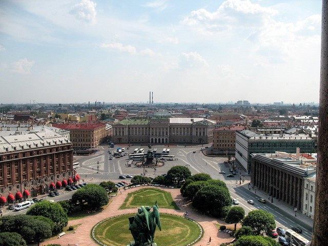
Computer technology - the Creed of today: The Barcelona Supercomputing Centre (with 'Mare Nostrum') located in a former chapel. Source.


So, while focus is still on the trillions of bacteria we have in our gut, we hope that it won't be long before common eukaryotic components of the intestinal microbiome will be studied and analysed alongside with bacterial communities. It says on Wikipedia that targeted studies of eukaryotic and viral communities are limited and subject to the challenge of excluding host DNA from amplification and the reduced eukaryotic and viral biomass in the human microbiome. Excluding host DNA is challenging, but not impossible, and who has actually documented that eukaryotic biomass in the human microbiome is 'reduced'?

The meeting was very well organised and took place at the Sheraton Hotel in Lisbon. I've storified a list of the #CMHH tweets here in case you are interested in more 'headlines'. I apologise for any misquotes.

Further reading:

Koren O, Knights D, Gonzalez A, Waldron L, Segata N, Knight R, Huttenhower C, & Ley RE (2013). A guide to enterotypes across the human body: meta-analysis of microbial community structures in human microbiome datasets. PLoS Computational Biology, 9 (1) PMID: 23326225

Andersen LO, Vedel Nielsen H, & Stensvold CR (2013). Waiting for the human intestinal Eukaryotome. The ISME Journal PMID: 23407309

Ivanov II, & Honda K (2012). Intestinal commensal microbes as immune modulators. Cell Host & Microbe, 12 (4), 496-508 PMID: 23084918

Brown J, de Vos WM, Distefano PS, Doré J, Huttenhower C, Knight R, Lawley TD, Raes J, & Turnbaugh P (2013). Translating the human microbiome. Nature Biotechnology, 31 (4), 304-8 PMID: 23563424

Blaser M, Bork P, Fraser C, Knight R, & Wang J (2013). The microbiome explored: recent insights and future challenges. Nature Reviews. Microbiology, 11 (3), 213-7 PMID: 23377500

Friday, May 10, 2013

Cell Symposium: Microbiome & Host Health - Lisbon 2013

My colleagues from Statens Serum Institut and I are heading to Lisbon, Portugal, tomorrow morning to attend the Cell Symposium on Microbiome and Host Health (link may be really busy now).

We are bringing a poster displaying some of our work related to our GUT18S project: A Novel Approach For Eukaryotic Phylogenetic Interrogation Of Clinical Samples Using Next Generation Sequencing Of SSU rRNA Genes; a pdf version of the poster can be downloaded here.

The GUT18S work is partly funded by the Marie Curie Actions (FP7) program.

Thursday, May 9, 2013

YouTube Video on Blastocystis Subtyping

For those who want to venture into Blastocystis subtyping - the easy way - I've recorded and uploaded a video on YouTube fyi.


For even more information, please visit a selection of relevant blog posts here.

Sunday, May 5, 2013

More on 'Bugs as Drugs'

This morning, I was doing a lazy ramble through my favourite blogs and found a post by Carl Zimmer on 'Bugs as Drugs' - primarily on probiotics. And I just came to realise that there is a very interesting tendency these years of using bugs as drugs in a variety of fields.

We are all very much aware of the worries about the increase in antibiotic resistance in bacterial and other pathogens. Moreover, it appears that sometimes antibiotic treatment leads to imbalance in the intestinal microbiota (dysbiosis); a well-known example is intractable Clostridium difficile infections which can potentially lead to pseudomembranous colitis.

C. difficile infection can lead to pseudomembranous colitis
Earlier this year, an article appeared in the renowned The New England Journal of Medicine on a randomised, controlled treatment study on duodenal infusion of donor faeces for recurrent C. difficile. The researchers found that the infusion of donor faeces was significantly more effective for the treatment of recurrent C. difficile infection than the use of vancomycin, the drug usually recommended in this situation. In fact 15/16 patients had resolution of C. difficile-associated diarrhoea upon first or second infusion; however, it might be worthwhile 'shopping around' for the right donor.

And so, how are these faecal transplants developed and administered? Well, it appears that donors are volunteers who have been through a selection process based on a questionnaire on risk factors of infectious diseases. Then donor faeces is screened for parasites (including Blastocystis and Dientamoeba - yes, it warms my heart to see this so explicitly spelled out in the paper... but I wonder which methods were used - it doesn't say!) and enteropathogenic bacteria. Moreover, blood samples from donors are screened for e.g. HIV, hepatitis and antibodies against e.g. Entamoeba histolytica and Strongyloides. Next, a donor pool is created with repeated screening every 4 months. On the day of infusion, faeces is collected by the donor and immediately brought to the hospital, where it is diluted with 500 mL of sterile saline. The solution is stirred, and the supernatant strained and poured in a sterile bottle. Within 6 h after collection of the faecal sample by the donor, the solution is infused through a nasoduodenal tube (2 to 3 mintues per 50 mL). Patients are subsequently monitored for 2 h. Apparently, this is how it works!

Monday, April 29, 2013

Transcriptomic Analysis of Blastocystis ST1!

BLASTing Breaking News!

Probably to support their genomic data, researchers in Andrew Roger's group in Canada have performed transcriptomic analysis of the Nand II strain, which belongs to Blastocystis sp. ST1.

Running from April 29 to May 2 is the SMBE (Society for Molecular Biology and Evolution) satellite meeting on Eukaryotic-Omics; the abstract booklet can be downloaded here. And fellow tweeps, don't let yourselves down by not following #SMBEeuks!

Until now, we've only known of one complete Blastocystis nuclear genome, namely that of ST7. Now, the release of the ST1 genome may be imminent! In any case, Roger's group have used their transcriptomic data to compare the protein content in ST1 with that in ST7, and it appears from their conference abstract that "the genes encoded by the Nand II strain (ST1) are surprisingly distantly related to ST7 orthologues, sharing on average ~50% identity at the protein level." And more: "Preliminary analyses allowed us to detect ~1000 genes in ST1 that have no homologue in Blastocystis sp. ST7". This means that the extreme genetic diversity that we see across the SSU ribosomal RNA genes is reflected and may be even more pronounced at nuclear genome level.

The group also studied genes acquired by lateral gene transfer (LGT; see previous post for more on LGT, also known as horizontal gene transfer), and what they basically found was that ST1 appears to have acquired bacterial genes related mainly to metabolism, while genes acquired from eukaryotes code for proteins related to cellular processes and signaling mechanisms.

Finally, they have discovered genes obtained by LGT that has had importance for Blastocystis' adaptation to parasitism; among these genes that enable resistance to host immune responses.

Roger's group is based at the Dept. of Biochemistry and Molecular Biology, Dalhouise University, Halifax, Nova Scotia in Canada.


'Invasive Blastocystis' in ECCMID 2013

ECCMID - the annual European Congress of Clinical Microbiology and Infectious Diseases (hosted by ESCMID) is currently taking place in Berlin. This year, I'm not attending, but I've been scanning the abstract book for 'Blastocystis', and it appears that an oral presentation was scheduled for yesterday in the "Emerging Infectious Diseases" section:

First of all: it's great to see fellow researchers screening larger (i.e. hundreds) of faecal DNAs by PCR for Blastocystis. I wish more people would do that to produce reliable data on prevalence and subtypes.

Now, as I've already mentioned, there are currently mainly two methods in use for subtyping, barcoding and STS PCR, and recently I evaluated these. To cut a long story short, barcoding is recommended for subtyping, since the STS method, which was used in the study by Tarasova et al. (abstract), appears to miss the majority of ST4 strains (the major genotype), and moreover, no STS primers exist for ST8 and ST9 (or any of the other 8 subtypes identified to date, but which have only been found in animals). So, the subtype data found in this study should be interpreted with this in mind.

Importantly however, I'm not sure whether the authors used the original Yoshikawa STS terminology or the terminology acknowledged in our 2007 consensus.

First, let us assume that consensus terminology is used. Then it's surprising to find ST5 in human samples in the first place, and finding a ST5 prevalence of 45% in a cohort of humans included in a larger study like this is very unlikely based on current evidence of more than 3,000 observations from all over the world, where the overall prevalence of ST5 in humans is <1%. Also, finding so much ST6 is also really striking. Also, if the consensus terminology is used, then I'm a bit puzzled why the authors put emphasis on ST7 not being found, since ST7 is relatively rare in humans.

And so let us assume that consensus terminology was not used, and the original Yoshikawa terminology was used instead. This would translate into STs 4, 6, and 7 not being detected in the CVH group. Which makes sense, since ST6 is extremely rare (at least in Europe), ST7 is only seen on occasion, and, as I said, the majority of ST4 infections are likely to go undetected by the STS method. However, ST4 appears quite common in Europe, and I suspect that it should be quite common in St Petersburg as well. But then there is one thing that comes to my mind: If ST4 infections are common, then there should be a relatively large number of samples detected by PCR which were untypable by PCR...and there is no information on untypable positive samples in the abstract...
But what is more:  STS subtype 5 translates into ST2 in consensus terminology, and similarly STS subtype 6 equals ST5 (yes, it may seem confusing, but we have provided a table in the 2007 consensus paper to make this easy). This means that no matter which of the two terminologies were used, ST5 is seen in abundance in patients with CVH in St Petersburg! Which is a very remarkable observation, and maybe more interesting than the rest of the data, which  I, by the way, find a bit difficult to follow (I expected to learn something about Blastocystis invasion, when I read the title of the abstract, but there is no data or information on invasiveness... and I'm very curious as to how the authors managed to obtain such a high number of samples from 'healthy people'! To evaluate the prevalence of Blastocystis in the control group, demographic data are needed, and a prevalence as low as 5.3% among healthy individuals makes me suspect that this control group consisted of newborns/toddlers who generally have a low prevalence of Blastocystis). Also, since when was ST1 'zoonotic'?

Anyway, often conference abstract are previews of upcoming articles, and so I expect that there will be a paper out soon from this group, and hopefully these issues will be clarified. The occasional confusion in Blastocystis epidemiology could be reduced to a minimum if everyone got into using barcoding and the Blastocystis 18S subtyping site (and go here for a video introduction to Blastocystis subtyping).

Are some citizens of St Petersburg infected by Blastocystis sp. ST5, a subtype seen primarily in livestock and African apes? Source

References:
Tarasova E, Suvorova M, Sigidaev A, Suvorov A. Blastocystis invasion in patients with chronic viral hepatitis in Saint Petersburg. ECCMID 2013 abstract O338.

Alfellani MA, Stensvold CR, Vidal-Lapiedra A, Onuoha ES, Fagbenro-Beyioku AF, & Clark CG (2013). Variable geographic distribution of Blastocystis subtypes and its potential implications. Acta Tropica, 126 (1), 11-8 PMID: 23290980

Stensvold CR (2013). Comparison of sequencing (barcode region) and sequence-tagged-site PCR for Blastocystis subtyping. Journal of Clinical Microbiology, 51 (1), 190-4 PMID: 23115257

Stensvold CR, Suresh GK, Tan KS, Thompson RC, Traub RJ, Viscogliosi E, Yoshikawa H, & Clark CG (2007). Terminology for Blastocystis subtypes--a consensus. Trends in Parasitology, 23 (3), 93-6 PMID: 17241816

Friday, April 26, 2013

This Month in Blastocystis Research (APR 2013)

I've been extremely bored all day writing up my evaluation of a (not so interesting) PhD thesis, and I thought I'd spice up my day by introducing a new series of posts on this blog inspired by so many other blogs, namely: This Month in Blastocystis Research! A place for me to go through some of the most recent papers on Blastocystis.

There is paper out by Gould and Boorom who look at the stability of Blastocystis surface antigen over time. They show that detection of Blastocystis by an immunofluorescense assay (IFA) is not hampered after1 year of storage of faecal material in formalin compared to results immediately after the sampling point. Detection of Blastocystis by IFA is something that is not often used (that's my impression, anyway), but makes sense in cases where laboratory analyses can be performed only weeks-months after sample collection (e.g. during field work), in which case samples need to be preserved. We usually, however, recommend storing faecal material in (70%) ethanol (in the relationship 1 part faecal sample to 4 parts of ethanol), where the sample is mixed with the ethanol initially by vortexing the tube (typically a 2 mL Eppendorf tube) for 5-10 min, and subsequently keeping the tubes away from light until further processing. Importantly, in contrast to formalin-fixed stool, ethanol-fixed stool can be made highly suitable for PCR by just washing the samples x3 in PBS prior to DNA extraction. An example of this methodology can be seen in our study of Blastocystis in members of the Tapirapé tribe in Mato Grosso, Brazil (go here for a free download).

I'd wish that Gould and Boorom had validated their findings by running a PCR on the samples too (specificity and sensitivity testing). The IFA assay was also used in a publication from 2010 by Dogruman-Al et al., who found a diagnostic sensitivity of the IFA assay of 86.7% compared to culture; also here, adding PCR would have been relevant to better determine the diagnostic qualities of the IFA assay.

I was lucky to be involved in field work in the Lao PDR in 2003 conducted by regional WHO authorities; preserving and analysing faecal samples for parasites by microscopy (Kato Katz) and - later - PCR was what we did!

Adding to the endless row of cross-sectional prevalence papers, there is an article out just now by Abdulsalam et al. (2013) on Prevalence, predictors and clinical significance of Blastocystis sp. in Sebha, Libya (free for download here). The study used culture (Jones' medium) as diagnostic modality and confirmed the existence of frequent asymptomatic carriage. The authors used questionnaire info and multivariate statistical analysis to identify risk factors for Blastocystis carriage among 380 individuals aged 1-75, and what I find really interesting is that they found that participants aged > 18 years were much more prone to having Blastocystis than participants < 18 years (P < 0.001). This is something that we see in Denmark too, and I'm currently trying to collect "sufficient proof"! Whether this is an age accumulation effect due to the chronicity of colonisation remains to be investigated. The authors also found that carriers were more likely to experience symptoms than those who were not carriers (P < 0.001), mainly abdominal pain (P < 0.001), but notably not diarrhoea (P = 0.117).
It's a pity that molecular data was not included the study from Libya. Incidentally, our group recently published subtype data from Sebha, Libya, and it appears that Blastocystis found in humans in Libya mainly belongs to ST1, whereas ST3 is often the most common subtype in most other countries, and what is more: ST4 appears virtually absent in Libya and the rest of Africa... But let's see: The investigators might have more data up their sleeve waiting to be published...

May I also again draw your attention to our recent paper on Blastocystis in non-human primates, in which we find that despite the fact that there is a great overlap of subtypes in human and non-human primates, it appears that ST1 and ST3 strains found in non-human primates differ genetically from those found in humans, indicating cryptic host specificity. We have additional data supporting the theory that Blastocystis in humans is a result of human-to-human transmission (anthroponotic) rather than animal-to-human (zoonotic) transmission. Which is really interesting, since the theory of zoonotic transmission of Blastocystis has been heavily (I dare not say purported, so I'll say) propagated. Having said that, I think we still need to look much deeper into barcoding of Blastocystis from pets and other synanthropic animals before we can make more poignant conclusions.

And, finally, yet another add for our recent review on Recent Development in Blastocystis Research!

Please note that I'm happy to take suggestions for future posts, and I'd also like to encourage guest blogging!

Suggested reading:

Abdulsalam AM, Ithoi I, Al-Mekhlafi HM, Khan AH, Ahmed A, Surin J, & Mak JW (2013). Prevalence, predictors and clinical significance of Blastocystis sp. in Sebha, Libya. Parasites & Vectors, 6 PMID: 23566585

Alfellani MA, Jacob AS, Perea NO, Krecek RC, Taner-Mulla D, Verweij JJ, Levecke B, Tannich E, Clark CG, & Stensvold CR (2013). Diversity and distribution of Blastocystis sp. subtypes in non-human primates. Parasitology, 1-6 PMID: 23561720

Alfellani MA, Stensvold CR, Vidal-Lapiedra A, Onuoha ES, Fagbenro-Beyioku AF, & Clark CG (2013). Variable geographic distribution of Blastocystis subtypes and its potential implications. Acta Tropica, 126 (1), 11-8 PMID: 23290980

Clark CG, van der Giezen M, Alfellani MA, & Stensvold CR (2013). Recent developments in Blastocystis research. Advances in Parasitology, 82, 1-32 PMID: 23548084

Dogruman-Al F, Simsek Z, Boorom K, Ekici E, Sahin M, Tuncer C, Kustimur S, & Altinbas A (2010). Comparison of methods for detection of Blastocystis infection in routinely submitted stool samples, and also in IBS/IBD Patients in Ankara, Turkey. PloS One, 5 (11) PMID: 21124983 

Gould R, & Boorom K (2013). Blastocystis surface antigen is stable in chemically preserved stool samples for at least 1 year. Parasitology research PMID: 23609598

Malheiros AF, Stensvold CR, Clark CG, Braga GB, & Shaw JJ (2011). Short report: Molecular characterization of Blastocystis obtained from members of the indigenous Tapirapé ethnic group from the Brazilian Amazon region, Brazil. The American Journal of Tropical Medicine and Hygiene, 85 (6), 1050-3 PMID: 22144442

Monday, April 15, 2013

Recent Developments in Blastocystis Research

I would like to draw your attention to an open access link to our 2013 review on Recent Developments in Blastocystis Research; please go here to read/download the paper.

Here's the abstract:

Blastocystis is a common parasite of the human large intestine but has an uncertain role in disease. In this review, we appraise the published evidence addressing this and its weaknesses. Genetic diversity studies have led to the identification of numerous subtypes (STs) within the genus Blastocystis and, recently, methods for studying variation within STs have been developed, with implications for our understanding of host specificity. The geographic distribution of STs is summarised and the impact this may have on investigations into the role of the organism in disease is discussed. Finally, we describe the organelle and nuclear genome characteristics and look to future developments in the field.


Wednesday, April 10, 2013

Blastocystis Hits The 1,000 Entry Mark In PubMed

Yesterday, the number of Blastocystis entries in PubMed reached 1,000! PubMed is a public resource comprising more than 22 million citations for biomedical literature from MEDLINE, life science journals, and online books.

In comparison, there are currently 7,641 entries on Entamoeba, 6,630 on Cryptosporidium and 235 entries on Dientamoeba.


I plan to introduce the "Hall of Fame in Blastocystis Research" in a future post, but already now I can reveal that the researcher with most Blastocystis-related publications is Dr Hisao Yoshikawa according to Web of Science (WoS), which currently returns 895 hits on a search on Blastocystis; Dr Yoshikawa has at least 43 publications on Blastocystis alone (WoS), and at least 37 Blastocystis-specific peer-reviewed journal articles (PubMed) since 1987.

Tuesday, April 9, 2013

Blastocystis in Non-Human Primates

If my recent blog post "Blastocystis aux Enfers" could be described as "Blastocystis meets Dante Alighieri", then this post might come across as "Blastocystis meets Sir David Attenborough" (with all due respect to both of these gentlemen!).

Non-human primates (NHPs) include apes (hominoids), Old World monkeys (cercopithecoids), New World Monkeys (ceboids) and prosimians such as lemurs. I have been so fortunate to be involved in a study of Blastocystis in NHPs; a study which was led by Dr Alfellani with several co-investigators, and which has just appeared online in the journal Parasitology (click here to be diverted to the the website - first view article section).

The study is the first of its kind aiming to provide a substantial insight into the host specificity of Blastocystis in NHPs and included subtype observations for 441 captive and free-living animals representing no less than 30 genera; most of the data were generated during the study, while sporadic observations from similar studies were also included.

It was a huge study with a lot of interesting information, and I will try and summarise some of the points here.

Apes such as bonobos, chimps, gorillas and orangutans were colonised by some of the most common subtypes in humans, namely ST1, ST2, and ST3, accounting for about 77% of the cases. Contrary to humans though, ST5 also appeared rather common, accounting for about almost 14% of the cases, and some of the gibbons studied had ST8. Interestingly, a chimp and a gibbon were found to be hosts of a new subtype, ST15.

Old World monkeys were studied to an even larger extent, and again, ST1, ST3 and ST2 predominated, accounting for about 95% of all cases of single subtype infection. Here ST5 was also seen (2%) but only in langurs/lutungs and vervet monkeys. Interestingly, ST8 was seen only in 1/226 cases. ST13 was found by colleagues in Tanzanian colobus monkeys (Petrasova et al., 2011), and 8% of the 226 cases represented mixed/unknown subtype infections.

Woolly monkey (Lagothrix lagotricha) (Source)

New World monkeys included in the study were mainly represented by woolly monkeys, and these were colonised first and foremost by ST8 (49%), but ST3, ST2, ST1 were also found. So was a single case of ST4, which in general appears to be surprisingly rare among NHPs.

A few observations on lemurs were included, and such animals appear to host a vast variety of subtypes with no particular predilection, hence ST1, ST2, ST4, ST8, ST10 and ST15.

Ring-tailed lemur (Lemur catta) (Source).

The most striking differences between humans and NHPs in terms of colonisation by Blastocystis subtypes is that humans are very rarely colonised by ST5, while this subtype appears common in apes and Old World monkeys. ST8 was seen only in arboreal apes and in woolly and howler monkeys, which are also tree-dwellers, and it is tempting to think that ST8 is found mainly in tree-dwelling NHPs; to my knowledge, ST8 has not been found in non-primate hosts, except for once in a bird. Human colonisation by ST8 has been demonstrated only very rarely, for instance in a Danish woman returning from holiday in Indonesia and in animal keepers. Conversely, ST4 is seen extremely rarely in NHPs, while very common in humans in some parts of the world, apparently especially in Europe. These clear discrepancies in subtype distribution in humans and NHPs may boil down to host specificity and/or apparent geographically restricted range of some subtypes.

Another striking observation was that cryptic host specificity exists in ST1 and ST3, meaning that ST1 and ST3 strains found in NHPs overall differ genetically from strains found in humans belonging to the same subtypes, adding support to our previous findings.This suggests that humans are generally colonised by other strains than those found in NHPs. It will be interesting to see, whether other types of hosts sharing these subtypes carry distinct, host-specific strains. While MLST is probably the best way of testing for this, a lot of information can be obtained simply by barcoding. Pets, for instance, may share subtypes seen in humans, and so barcoding of "pet blasto" may be one of the very interesting pathways to knowledge.

We found no evidence of those subtypes that we have nicknamed "avian subtypes", namely ST6 and ST7. In some parts of the world, these two subtypes do not appear uncommon in humans; in Denmark and Sweden, for instance, ST7 is seen on quite a few occasions. But, interestingly, both STs are apparently absent in NHPs.

Langurs - the front cover of one of my favourite books showcasing works by the magnificent Walton Ford.

Incidentally, there is a sequence in GenBank from a gorilla (JX159284) which possibly represents a novel subtype, which is related to reptilian Blastocystis, and so it appears that the host spectrum and diversity of Blastocystis in NHPs continues to unfold.

A recent study saw that faecal microbiomes of wild non-human primates co-vary with host species, hence reflecting host phylogeny. This was evidenced by higher intra-species similarity among wild primate species, which may reflect species specificity of the microbiome in addition to dietary influences. This may in part explain the differences in Blastocystis subtypes seen in different NHP host species, but it is also possible that differences in subtypes reflect differences in habitat (and thereby possibly exposure) or geographical differences in subtype distribution. Indeed Homo sapiens is host to a variety of subtypes, and while ST4 is common in Europe, it appears virtually absent in many other parts of the world. Likewise, the differences in the prevalence of ST8 may reflect differences in geographical distribution, habitat and diet (arboreal vs. ground) as well differences in host specificity.

The overall interesting thing here is the schism of exposure vs. host specificity.


Suggested reading:

Alfellani, M., Jacob, A., Perea, N., Krecek, R., Taner-Mulla, D., Verweij, J., Levecke, B., Tannich, E., Clark, C., & Stensvold, C. (2013). Diversity and distribution of Blastocystis sp. subtypes in non-human primates Parasitology, 1-6 DOI: 10.1017/S0031182013000255

Yildirim S, Yeoman CJ, Sipos M, Torralba M, Wilson BA, Goldberg TL, Stumpf RM, Leigh SR, White BA, & Nelson KE (2010). Characterization of the fecal microbiome from non-human wild primates reveals species specific microbial communities. PloS one, 5 (11) PMID: 21103066

Stensvold CR, Arendrup MC, Nielsen HV, Bada A, & Thorsen S (2008). Symptomatic infection with Blastocystis sp. subtype 8 successfully treated with trimethoprim-sulfamethoxazole. Annals of tropical medicine and parasitology, 102 (3), 271-4 PMID: 18348782

Stensvold CR, Alfellani M, & Clark CG (2012). Levels of genetic diversity vary dramatically between Blastocystis subtypes. Infection, genetics and evolution : journal of molecular epidemiology and evolutionary genetics in infectious diseases, 12 (2), 263-73 PMID: 22116021

Saturday, April 6, 2013

Technical issues...

I'm experiencing a problem with my post titles being displayed as uncials (sort of nested in each post with blog text wrapped around it), when using Firefox or Chrome (also the headline link function has been disabled). I'm trying to fix it, but for now, I recommend viewing the blog in Internet Explorer rather than eg. Firefox or Chrome. If anyone knows how to fix this, please give me a shout!

Have a great weekend!

Wednesday, April 3, 2013

Blastocystis and IBD

We recently published what could be seen as a pilot study on inflammatory bowel disease (IBD) and the two most common intestinal parasites, Blastocystis and Dientamoeba fragilis.

The aim of the study was to identify possible differences in the prevalence of infection with Blastocystis and D. fragilis in patients with active and inactive IBD compared to controls.

We included 100 Danish patients with IBD (42 with Crohn's Disease, 41 with ulcerative colitis and 17 with ileal pouch-anal anastomosis) and 96 controls, used state-of-the-art diagnostics for Blastocystis and D. fragilis (PCR) and we saw striking differences in prevalence. While 19% of all healthy individuals had Blastocystis, only 5% of those with IBD had Blastocystis, and of the 42 patients with Crohn's Disease, only 1 had Blastocystis. In contrast, D. fragilis was not more common in healthy individuals than in IBD patients. Also, in patients with ulcerative colitis, Blastocystis was significantly more common in patients with inactive disease compared to patients with active disease.

Absence of Blastocystis in patients with Crohn's Disease and active ulcerative colitis may be due to unfavourable conditions for colonisation and should be explored further in order to investigate whether these potentially unfavourable conditions reflect differences in the composition of the microbiota in these patients, and/or whether this has something to do with host immunity. We are currently confirming the virtual absence of Blastocystis in Crohn's patients in another study based on metagenomic analysis of faecal DNA, and it will be very interesting to analyse the differences in Blastocystis prevalence in view of potential differences in bacterial communities.

The literature on Blastocystis and IBD is relatively limited, and I plan to return, maybe later this year, with a more elaborate post on the topic.

Reference:

Petersen AM, Stensvold CR, Mirsepasi H, Engberg J, Friis-Møller A, Porsbo LJ, Hammerum AM, Nordgaard-Lassen I, Nielsen HV, & Krogfelt KA (2013). Active ulcerative colitis associated with low prevalence of Blastocystis and Dientamoeba fragilis infection. Scandinavian journal of gastroenterology PMID: 23528075

Friday, March 29, 2013

Happy Birthday!

In these very hours, my daughter is turning two years old! This blog was put up one year ago as she was celebrating the conclusion of her first year on this planet, and given all the fun I've had along the way putting up posts on this and that, I'd like to dedicate the blog to her. In return(!), I take the liberty of using some of her artwork for this post which marks the birthday of the Blastocystis Parasite Blog.

Artwork by Raiya Rochelle Traub

It's surprising to me that this blog has had more than 50,000 views in only one year. Due to all the feedback I get, I'm prone to believe that most of the page views reflect factual "blog consumption" (rather than referral spam and bots). Anyway, even if there were only a few people out there who'd stop by every now and then, my efforts would certainly be worthwhile. 
Blastocystis has been known for more than 100 years. But it is only recently that we have found tools to enable accurate distinction of Blastocystis carriers from non-carriers, thanks to DNA-based diagnostic methods. Last year, we published a paper on our new real-time PCR in Journal of Clinical Microbiology, and it seems as if we now finally have the chance to try and use it for screening a larger panel of faecal DNAs from patients with and without intestinal symptoms to get an idea about the factual prevalence of Blastocystis in this type of samples with the added benefit of analysis of colonisation intensity. It's very exciting...

And to those who are involved in Blastocystis subtyping, -  in case you didn't see it, there is a paper out on the comparison of the two principal methods used for subtyping which you might find useful.
I've also added a few lines on barcoding in "Lab Stuff" for those who are new to subtyping and want to practice a bit - please go here.

We are currently trying to strengthen collaborative efforts of different labs across the world and we are facing some very exciting challenges, involving the generation and analysis of data output related to genomics, transcriptomics, metagenomics and possibly proteomics; more about that in "Season II" of the Blastocystis Parasite Blog!

But for now: Happy birthday, Raiya! And Happy Easter everyone!

Suggested reading:

Stensvold CR (2013). Comparison of sequencing (barcode region) and sequence-tagged-site PCR for Blastocystis subtyping. Journal of Clinical Microbiology, 51 (1), 190-4. PMID: 23115257 

Stensvold, C., Ahmed, U., Andersen, L., & Nielsen, H. (2012). Development and Evaluation of a Genus-Specific, Probe-Based, Internal-Process-Controlled Real-Time PCR Assay for Sensitive and Specific Detection of Blastocystis spp. Journal of Clinical Microbiology, 50 (6), 1847-51. DOI: 10.1128/JCM.00007-12

Thursday, March 21, 2013

LUMINEX xMAP Technology in Parasite Diagnostics

Over the past few years nucleic acid based methods have revolutionised parasite diagnostics in modern clinical microbiology (CM) labs. Real-time PCR is really gaining a foothold in CM labs, but despite the opportunity for plexing, mostly only up to 6 DNA targets can be included in each assay (due to the number of available channels).

LUMINEX xMAP technology used for detection of specific nucleic acids (Dunbar, 2006) bypasses this limit, and up to 100 DNA targets can be included in one single assay in a 96-well plate format. You can read about the technology here.


Thursday, March 14, 2013

Extremophilic Eukaryotes

My recent post Blastocystis aux Enfers was my "literary take" on biological adaptation of intestinal parasitic protists, using Blastocystis as an example. As a parasitologist you'd come across many peculiar and shrewd biological adaptations and life cycles, and I hope to be able to give some examples in a future post. Actually, there is a parasite which is quite common in humans, maybe even just as common as Blastocystis, which is also single-celled, but which may have a much more complicated life cycle than Blastocystis, namely Dientamoeba fragilis; a colleague of mine is currently doing his PhD on Dientamoeba and he has collected multiple sources of evidence to confirm the hypothesis that this parasite is transmitted by a vector, namely pinworm, probably along the same way that Histomonas meleagridis – the cause of blackhead disease in especially turkeys – is transmitted by heterakids (which again are transmitted by parathenic hosts such as earthworms, which get eaten by turkeys, chickens, etc.). Anyway, I’ll probably get back to Dientamoeba, once his data are out.

Meanwhile, Blastocystis comes out of a very heterogeneous group of organisms called Stramenopiles, many of which are algae. Algae are photosynthetic organisms found in habitats as diverse as glacial ice and hot springs.One of these algae is named Galdieria sulphuraria, which is a remarkable unicellular eukaryote inhabiting hostile environments such as volcanic hot sulfur springs where it is responsible for about 90% of the biomass; indeed this certainly qualifies as "Galdieria aux enfers"!

Tuesday, March 12, 2013

Blastocystis video

Just saw this on YouTube and had to share it. This is Blastocystis (and other microorganisms) viewed through a microscopy (light microscopy). Note that this is Blastocystis from a chicken, but Blastocystis from humans looks the same; at least I don't know how to tell the difference. I wonder whether this is from a culture or a completely fresh egestion... looks more like a culture to me. Note how the Blastocystis looks almost like fat cells...

The video comes with some nice music as well!


Wednesday, March 6, 2013

Open Access papers in Nature Reviews on functional dyspepsia

"Functional dyspepsia is one of the most common functional gastrointestinal disorders worldwide. Although the condition does not affect life expectancy, it can have a marked influence on quality of life, and is associated with a high economic burden; an estimated US$1 billion per year is spent on the management of functional dyspepsia in the USA alone. This comprehensive Focus issue from Nature Reviews Gastroenterology & Hepatology contains seven Reviews that have been specially commissioned to cover key themes in functional dyspepsia. Experts from around the world provide up-to-date overviews of the most important topics in the field, including the influence of dietary, lifestyle and psychosocial factors, relevance of Helicobacter pylori infection, overlap with GERD, changes in gastrointestinal tract structure and function, symptom pattern and validity of the Rome III criteria, as well as current and emerging treatment options."

For the bunch of papers, please go here.

Saturday, February 23, 2013

Blastocystis aux Enfers

We tremble at the thought of being devoured by a ferocious animal, - of ending our days in a narrow, suffocating slimy tube covered in acidic, nauseating glaze! Remarkably, for some eukaryotic beings, this is the only way forward if they want to carry on with their lives! Intestinal protists such as Blastocystis are in a state of hibernation when outside our bodies and the only thing that may rouse these Sleeping Beauties to action is the passage through low pH enzyme ponds. They thrive, grow and raise their progeny only in the swampy Tartarus of our large intestines; they bequeath to their offspring the affinity for this gloomy, filthy slew; this murky, densely populated, polluted channel, and when the pool of poo becomes all too arid, they know it’s time to buckle up, shut down, and prepare themselves for the great unknown which can potentially mean death to them if eventually they are not lucky enough to be gulped down by another suitable host.

Source
And yet, despite their remarkable modesty and humble requirements these little buggers are being bullied by their inhospitable human hosts; we’d throw anything at them to force them out, organic and inorganic compounds meant to arrest or even kill them. But the whelps of Blastocystis appear extremely resilient, which may hold the key to part of their success; they stay afloat on the Styx of our bowels. In order to eschew Flagyl, perhaps they bribed Phlegyas?

I think it's sometimes useful to put things into a completely different perspective. In any event, from an evolutionary biology standpoint it is highly interesting that a genus which is genetically related to water molds such as those causing potato blight and sudden oak death, has so successfully adapted to a parasitic, anaerobic life style, capable of protractedly colonising a plethora of very diverse host species including members of primates, other mammals, birds, reptiles, amphibians and arthropods and thereby evading innate and adaptive immune defenses from such a diverse range of hosts. One could be inclined to say: Well done! But which is it? Parasitism? Commensalism? Mutalism? Symbiosis? And what will happen to Blastocystis in the future? Will this successful crusader eventually succumb to our avid but maybe imprudent war strategies? And if so, what will happen to us after removing such a common player from our intestinal ecosystems?

Friday, February 22, 2013

Bubbly Blasto!

Yesterday, I was checking up on a fresh Blastocystis culture. I loaded 20 µL of the culture "sediment" on to a glass slide, placed the cover slip on top and examined it by light microscopy. While examining the slide, I observed a multitude of dividing cells, indicating vigorous growth and a thriving strain, and once again I was struck by the appearance of dividing Blastocystis. This is basically what they may look like:

Like soap bubbles really, only a lot smaller obviously (mikrons), and somewhat opaque! You'll see them in different sizes and the way they divide looks just like this. Apparently some sort of random budding or multiple fission. You'll see little more than this bubbly structure, which means that there are very few morphological hallmarks to describe. A few nuclei may be discernible along the cytoplasmatic rim, but that's about it when you use light microscopy. Ultrastructural and biochemical analysis is required if you hope to be able to describe some of the processes involved in reproduction.

We often say that Blastocystis organisms representing different subtypes are morphologically indistinguishable; what this actually means is that we do not have the tools to differentiate them morphologically. There may actually be great variation between strains in terms of for instance how they grow in vivo and in vitro and maybe also how they reproduce. Vacuolar forms are the most common form seen in xenic cultures, but other morphotypes are sometimes observed, for instance the granular stage, which, in my experience, is typically seen in cultures that are not “well looked after”, i.e. where medium is not being replaced about twice a week. Dunn and colleagues. (1989) observed that the granular stage could arise from vacuolar stages in cultures where the concentration of horse serum was increased.

I have previously stated that there is no evidence for phagocytosis in Blastocystis. Actually, Dunn et al. (1989) captured what they thought to be bacterial engulfment by ultra-structural analysis, and they also observed bacteria-engulfing pseudopodia in amoeboid stages, in which degraded bacteria were observed. I don't think that I've ever come across this amoeboid stage, but it has been described by quite a few researchers.

Anyway, let's hope for another kind of bubbles this Friday night!

Suggested reading:

Dunn LA, Boreham PF, & Stenzel DJ (1989). Ultrastructural variation of Blastocystis hominis stocks in culture. International Journal for Parasitology, 19 (1), 43-56 PMID: 2707962

Saturday, February 16, 2013

Waiting For The Human Intestinal Eukaryotome

We were lucky enough to have a paper accepted for publication in the ISME Journal (Nature Publishing Group) in which we call for data on the "human intestinal eukaryotome".

In the paper, we start out:

"Recent developments in Next Generation Sequencing (NGS) technologies have allowed culture-independent and deep molecular analysis of the microbial diversity in faecal samples, and have provided new insights into the bacterial composition of the distal gut microbiota. Studies of the microbiome in different patient groups using metagenomics or 16S rRNA gene sequencing are increasing our knowledge of how the microbiota influences health and disease. The majority of recent advances in our understanding of human microbiota structure and dynamic changes in disease were made through phylogenetic interrogation of small subunit (SSU) rRNA (Paliy and Agans 2012). However, until recently such studies have generally failed to include data on common eukaryotic, endobiotic organisms such as single-celled parasites and yeasts ('micro-eukaryotes'). This deficiency may strongly bias the interpretation of results and ignoring an entire kingdom of organisms is a major limitation of human microbiome studies."

Saturday, February 2, 2013

Blog Feedback

I'm very thankful for all the positive feedback I get from readers across the globe, mostly by email. Due to time limits I can only respond to 5-10% of the mail, and I'm sorry for not getting back to the rest of you.

Meanwhile, this blog currently holds more than 60 posts, and you will also find a lot of key words in the right side bar, so take your time and browse a few posts or look up a few relevant key words, -  you might find an answer to one or more  of your questions.

Having said that, I try to read all my email, and I am listening! The feedback and questions that I get are vital for our work and help us identify the avenues that we need to take to unveil the many mysteries of Blastocystis.

And let me just say this for now: A proper microbiological work-up (by state-of-the-art methods, including PCR for intestinal parasites), is something that is offered on a routine basis in only very few laboratories, and also the number of clinically orientated Blastocystis research centres can be counted on one hand, I believe. Subtyping of Blastocystis is currently done mostly in epidemiological surveys (as part of research projects), and I suspect that our lab is one of the very few labs in the world doing subtyping on a routine basis.

Oh, and I've been asked by some readers about how to get blog updates. It's easy: You can follow this blog by email, - just scroll down and find "follow by email" in the right side bar and enter your email address. You can also subscribe to posts via atom (go to the very bottom of the page).

And then here's a little something about stomach acidity and intestinal microbiota from Scientific American, - but make sure to read the comments underneath the post too!
 

Monday, January 14, 2013

A Penny For Your Thoughts

So, what should we do about Blastocystis? What do we want to know?

I believe the imminent answer to the latter question is easy: We want to know whether it’s pathogenic, whether we should treat it and how. But I also think that there are many other interesting aspects of Blastocystis which are also of broad interest to the general public, namely: How about the many cases of asymptomatic Blastocystis carriage? What does Blastocystis do in our guts? Could it have any potentially beneficial impact on our health?

Given the fact that Blastocystis has not been implicated in any outbreaks (admittedly: I guess that no one actually ever looked for Blastocystis in outbreak investigations... except for me!), I reckon that the chance of it being involved in acute diarrhoea is small. So, in that respect it's very different from the other intestinal protists such as Giardia, Cryptosporidium, Cyclospora, microsporidia, even Entamoeba histolytica. It's actually more reminiscent of helminth infections, which are are often chronic, and when light hardly give rise to symptoms (depending on species that is!).So I'm more thinking along the lines of co-evolution, adaptation, etc.

Maybe future research will call for a shift in paradigm, but until then I think that we should do what we already can, just at a larger scale and see where it takes us, namely:

Saturday, January 5, 2013

Where Are We On Blastocystis Subtypes?

As mentioned, Blastocystis exhibits remarkable intrageneric diversity, which is continuously being explored by us and our colleagues. We are convinced that the genus of Blastocystis comprises multiple species, but for now we call them "ribosomal lineages" or "subtypes" and allocate numbers to each subtype, hence ST1, ST2, etc. While the number of subtypes that can be found in humans remains stable, we and our colleagues are still expanding the subtype universe in non-human hosts (I will be blogging on this shortly).

Barcoding currently represents state-of-the-art in Blastocystis subtyping, and luckily this method appears to gain a foothold in labs across the world.

Nine subtypes have been found in humans, but some of them only on rare occasions. A recent study going out from London School of Hygiene and Tropical Medicine and led by Dr Alfellani and published just now in Acta Tropica looked at 356 Blastocystis sequences from samples from the UK and Libya, but also from sub-Saharan Africa, namely Liberia and Nigeria.